

Dermal Filler Vision Loss: What Patients Should Know About Vascular Occlusion and Safety in Abu Dhabi
Dermal fillers are widely used to restore facial volume and refine contours. Most adverse effects are mild and temporary, such as bruising, swelling or redness. However, the literature also describes rare but severe vascular complications, including skin necrosis and permanent vision loss. Understanding the anatomy, warning signs and importance of rapid escalation is a core part of safe filler practice.
MEDICAL SAFETY NOTE
This page is educational. Sudden loss or blurring of vision, severe eye pain, new double vision, ptosis, ophthalmoplegia, or neurological symptoms after filler injection require emergency medical assessment. Treatment decisions should be made by appropriately qualified emergency, ophthalmology and related specialists.
What is filler-associated vision loss?
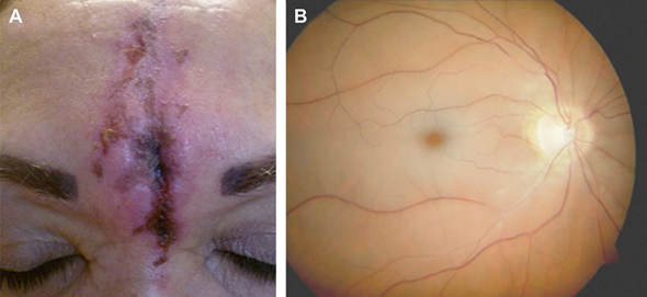
Filler-associated vision loss refers to visual impairment caused by vascular obstruction after cosmetic soft-tissue filler injection. The source review describes ocular complications ranging from branch retinal artery occlusion to central retinal artery occlusion and ophthalmic artery occlusion. The severity depends on the vessel involved and the degree and duration of ischemia.
The review emphasizes that blindness is rare but potentially irreversible. It also notes that severe events may occur alongside skin ischemia or necrosis because the same arterial network supplies the skin and has connections with the orbital circulation.
How can facial filler affect the eye?
The face contains an extensive network of arterial connections. The proposed mechanism is retrograde embolization: filler is inadvertently introduced into a facial artery, injection pressure pushes material backward through anastomoses, and normal arterial pressure may then carry the material forward into the ophthalmic artery or one of its branches.
This explains why a facial injection far from the eye can, in rare circumstances, affect the ocular circulation. The source specifically highlights connections involving the supratrochlear, supraorbital, angular, dorsal nasal, infraorbital and superficial temporal arterial systems.
Why “safe zones” are difficult to define
The review describes facial vascular anatomy as highly variable and richly anastomotic. For that reason, safety depends less on assuming that an area is completely risk-free and more on detailed anatomic knowledge, conservative technique, low injection pressure, small increments, careful plane selection and immediate recognition of complications.
High-risk anatomy discussed in the source
| Region | Why it matters |
|---|---|
| Glabella / lower forehead | The supratrochlear and supraorbital arteries are direct branches of the ophthalmic circulation and can lie close to common glabellar creases. |
| Nose / nasal sidewall | The angular and dorsal nasal arteries form important connections between facial and ophthalmic arterial systems. |
| Nasolabial fold | The facial/angular system and lateral nasal branches create pathways that can connect toward the ophthalmic circulation. |
| Temple | The superficial temporal system can connect with supraorbital or supratrochlear branches; vascular depth varies by plane. |
| Infraorbital region | The infraorbital artery exits near the infraorbital foramen and connects with other facial/orbital vessels. |
| Lips | The superior and inferior labial arteries have variable courses; the article emphasizes caution despite no clinical blindness cases in the reviewed evidence for lip augmentation. |
What types of ocular complications can occur?
- Ophthalmic artery occlusion — a proximal and particularly severe event affecting retinal and choroidal perfusion.
- Central retinal artery occlusion — can cause profound sudden visual loss and characteristic retinal ischemic findings.
- Branch retinal artery occlusion — visual deficit can vary according to the retinal territory involved.
- Ocular ischemic syndrome — may include hypotony, corneal edema, ophthalmoplegia and other signs of widespread orbital ischemia.
- Ischemic oculomotor palsy — may present with diplopia, ptosis or restricted eye movement.
- Cerebral infarction — rare reports describe filler emboli reaching intracranial circulation, making neurological symptoms an additional emergency concern.
Warning signs after filler that need immediate attention
EMERGENCY RED FLAGS
Seek urgent medical assessment for sudden vision loss or blurring, a new visual field defect, severe ocular or periocular pain, double vision, ptosis, abnormal eye movement, marked skin blanching or dusky discoloration, or new neurological symptoms.
The source review stresses early recognition, particularly when severe post-injection pain or visual symptoms are present. If an ocular vascular event is suspected, further filler injection should stop and ophthalmology referral should occur immediately.
Why timing matters
Retinal tissue has limited tolerance to ischemia. The review cites experimental work showing irreversible retinal damage can occur after a relatively short period of arterial occlusion. This is why suspected filler-related visual compromise should be managed as a time-critical emergency rather than observed for spontaneous improvement.
What does the source say about treatment?
The source review is clear that there is no reliably effective treatment for iatrogenic retinal embolism and that prompt ophthalmology evaluation is essential. Historical measures aimed at lowering intraocular pressure or dislodging embolic material have had inconsistent or disappointing results, and several experimental approaches have not demonstrated reliable visual recovery.
For hyaluronic-acid filler, hyaluronidase is important for managing accessible HA-related vascular compromise and should be available in practices that inject soft-tissue fillers. However, the review notes that retrobulbar or other attempts to deliver hyaluronidase to restore retinal perfusion have not shown consistently successful visual outcomes. Specialist-directed care remains essential.
IMPORTANT DISTINCTION
Hyaluronidase can dissolve hyaluronic acid filler, but the source does not support presenting it as a guaranteed treatment for established retinal or ophthalmic artery occlusion. Any eye-related symptoms need emergency specialist management.
Prevention principles highlighted in the literature
- Know the vascular anatomy and the expected depth of major vessels in each treatment area.
- Use small amounts per pass and inject slowly to reduce ejection pressure.
- Avoid large boluses and excessive injection force.
- If significant resistance is encountered, stop and reposition rather than increasing force.
- Keep the needle or cannula moving when using linear injection techniques rather than depositing a large static bolus.
- Remember that a cannula does not eliminate arterial injury risk; cannulas can penetrate vessels under certain conditions.
- Use region-specific technique and plane selection rather than one universal approach for the entire face.
- Be prepared to recognize and escalate a suspected vascular event immediately.
Needle versus cannula: is one completely safe?
No. The source notes that cannulas may reduce risk in some circumstances, particularly larger cannulas placed parallel to vessels, but cadaveric work shows that cannulas can still penetrate arteries. Needles can also enter vessels. The practical message is that instrument choice is only one part of risk reduction; anatomy, plane, pressure, volume, movement and clinical vigilance remain important.
Area-specific safety concepts from the source
| Area | Source-derived safety concept |
|---|---|
| Temple | Palpate, mark and avoid the superficial temporal artery; plane selection matters because vessels occupy different fascial levels. |
| Glabella / forehead | Arterial depth changes as the vessels ascend, so injection depth should reflect the anatomy rather than follow one rule everywhere. |
| Infraorbital area | The infraorbital foramen and artery are key landmarks; the article stresses avoiding direct risk near the foramen. |
| Nasolabial fold | The upper and lower portions have different arterial relationships; low-volume linear techniques and awareness of the angular/lateral nasal vessels are emphasized. |
| Lip | Use small controlled deposits and respect the variable position of the labial arteries, especially near the wet-dry junction. |
How should patients in Abu Dhabi think about filler safety?
When choosing dermal filler treatment in Abu Dhabi, safety should be part of the consultation — not an afterthought. A proper assessment should discuss the treatment area, the intended product, realistic goals, previous filler or surgery, relevant medical history, and the rare possibility of vascular complications. Patients should also know what symptoms require urgent contact or emergency assessment.
A strong filler consultation is not simply about selecting a brand or achieving a particular shape. It is also about anatomy, indication, conservative planning, complication readiness and clear aftercare communication.
Frequently asked questions
Can dermal filler cause blindness?
Yes, although it is rare. The mechanism described in the medical literature is accidental arterial entry of filler followed by embolization into the ophthalmic or retinal circulation.
Which filler areas are associated with higher eye-related vascular risk?
The source highlights the glabella, forehead, nose, nasolabial region, temple and infraorbital area because of their arterial connections with the ophthalmic circulation.
Is blindness after filler usually temporary?
It can be permanent. In the source review, ophthalmic artery occlusion had a particularly poor visual prognosis, and many reported severe cases did not regain vision.
What should I do if my vision changes after filler?
Stop the procedure and seek urgent ophthalmology or emergency medical assessment immediately. Sudden visual change after filler should not be watched at home.
Can hyaluronidase always reverse filler-related blindness?
No. Hyaluronidase can dissolve hyaluronic acid filler, but the source review reports that attempts to restore vision in established ophthalmic or retinal occlusion have not produced consistently successful outcomes.
Is a cannula safer than a needle for filler?
A cannula may lower risk in some settings, but it is not risk-free. The source describes situations in which cannulas can still penetrate arteries.
Does aspiration guarantee that the needle is not in a vessel?
No. The source warns that a negative aspiration result can create false reassurance because very small movements can change the position of the needle or cannula.
Why is the glabella considered a higher-risk area?
The supratrochlear and supraorbital arteries in this region are direct branches of the ophthalmic circulation, creating a potential route for embolization toward the eye.
Can filler-related vascular occlusion affect the brain?
Rare cases of cerebral infarction have been described when embolic material reaches intracranial circulation. New neurological symptoms after filler require emergency evaluation.
What is the safest filler approach?
There is no single technique that eliminates risk. The source emphasizes detailed anatomy, small volumes, slow low-pressure injection, appropriate plane selection, avoidance of large boluses, and immediate recognition of complications.
Considering dermal filler in Abu Dhabi?
A consultation should assess facial anatomy, treatment goals, prior procedures and the safest treatment plan for the specific area.
For sudden visual or neurological symptoms after filler, seek emergency medical care rather than booking a routine consultation.