
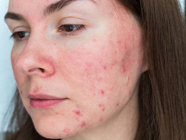
Expected Side Effect or True Complication ?
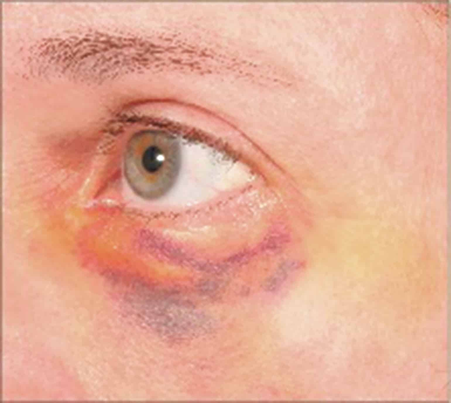
A major source of anxiety after injectable treatment is distinguishing a normal post-injection response from a complication. The source chapter notes that local tenderness, erythema, edema and ecchymosis are among the most common adverse events after filler and are usually mild, localized and transient.
The clinical pattern matters more than the mere presence of swelling or bruising. A small bruise that gradually improves is very different from worsening pain with blanching, a reticulated purple color change, progressive swelling with fever, or sudden visual symptoms.
| Pattern | Often expected / self-limited | Needs prompt medical review |
|---|---|---|
| Pain | Mild injection-site soreness that improves | Pain out of proportion, worsening pain, pain with skin color change or visual symptoms |
| Swelling | Localized edema that peaks and settles | Progressive, asymmetric, hot, tender, recurrent or delayed swelling |
| Bruising | Purple discoloration that behaves like a bruise | Blanching or dusky/reticulated discoloration suggesting reduced blood flow |
| Lumps | Small early swelling or product feel | Persistent visible papule, hard nodule, inflamed nodule or recurrent lesion |
| Vision | No visual change | Any visual loss, blur, field defect, double vision or ocular pain after filler |
Immediate vs Delayed Filler Complications
The chapter uses timing as a practical diagnostic framework. Acute complications develop up to approximately one week after treatment and include injection-site reactions, nodules, infection, hypersensitivity, tissue necrosis and blindness. Delayed complications may arise weeks to years later and include infection, biofilm-related problems and granuloma formation.
Can filler complications happen months or years laterYes. Delayed inflammatory reactions, biofilm-related inflammation and foreign-body granulomas can present long after the original filler treatment. A patient should tell the evaluating clinician what product was injected, where it was placed and approximately when it was injected, even if the treatment was years earlier.
Common Early Effects : Pain, Swelling and Bruising
Pain can result from tissue expansion during injection and from repeated needle or cannula entry. Lips and the perioral region are particularly sensitive because of dense sensory innervation. Swelling and bruising result from local tissue trauma and vascular permeability and usually resolve over several days.
The source stresses medication and supplement history before treatment. Importantly, medically necessary antiplatelet or anticoagulant therapy should not simply be stopped for an elective filler procedure; the risk-benefit decision belongs with the prescribing clinician.
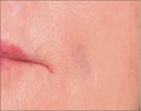
Tyndall Effect & Incorrect Filler Placement
When HA filler is injected too superficially, it may become visible as a blue-gray hue or discrete bump beneath thin skin. The chapter identifies this as the Tyndall effect. It is an example of a placement complication rather than an allergic reaction.
Management depends on the filler and the nature of the problem. The chapter notes that persistent HA papules or nodules related to superficial placement may be treated with hyaluronidase by a clinician. This is different from treating an infected or inflammatory nodule, where the diagnosis must be established first.
Filler Infection : Early and Delayed Presentations
Any injectable procedure breaches the skin barrier. The review describes bacterial infection as the dominant infectious complication, while fungal, viral and polymicrobial infections have also been reported. Early infection may present as tender erythematous nodules, cellulitis or abscess; delayed infection may be more indolent and can overlap clinically with biofilm or inflammatory nodules.
The source emphasizes prevention through antiseptic skin preparation and careful technique. Patients with immunocompromise, diabetes, chronic sinus or dental problems may need additional attention. Dental procedures and active infection should be discussed during planning because hematogenous seeding of filler material is a theoretical concern.
Patient Safety PointA painful, red, warm, fluctuant or progressively swollen injection site – especially with fever or malaise – should not be self-treated as “normal filler swelling.” It needs clinical assessment to distinguish infection, inflammatory reaction and other causes.
Herpes Reactivation After Filler
The chapter reports herpes reactivation within the first 24 to 48 hours after filler, particularly in the perioral region and nasolabial folds. A history of recurrent herpes should therefore be discussed before lip or perioral injections, and active outbreaks are a reason to delay elective treatment.
Delayed Inflammatory Reactions (DIRs)
Delayed inflammatory reactions can develop after both HA and non-HA fillers. The chapter describes discoloration or erythema, pain, nodules, induration, tissue hardening and solid edema as possible presentations. These reactions may occur months to years after treatment.
For HA fillers, the chapter discusses possible contributions from cross-linking technology, molecular-weight fragments and immune triggers. It also notes that infection must be considered before assuming that every delayed nodule is sterile inflammation.
Can Illness, Vaccination, Trauma or Dental Work Trigger Swelling Around Old Filler ?
The chapter describes viral or bacterial infections, vaccinations, facial trauma and dental work as potential immune triggers for delayed filler inflammation in susceptible patients. It also reviews rare reports of facial or lip swelling around previous HA filler during the COVID-19 era.
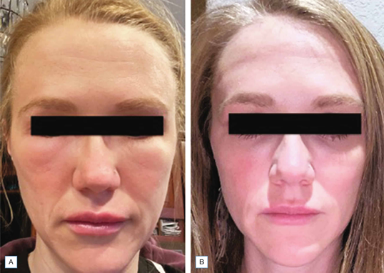
PLLA (Sculptra) Papules & Nodules
The source chapter explains that early experience with PLLA had a higher incidence of papules and nodules, much of which was linked to older preparation, hydration and placement techniques. Modern dilution and deeper placement recommendations reduced this problem, although delayed nodules can still occur.
Dynamic, thin-skinned regions such as the periocular and perioral areas require particular caution because product aggregation may be more visible or palpable. The broader lesson is that a biostimulatory filler has technique-specific risks that are different from those of HA.
Calcium Hydroxyapatite (CaHA / Radiesse) Nodules
CaHA can also form visible nodules if injected too superficially. The chapter highlights thin-skinned regions such as the tear trough as higher-risk for visible product irregularity. It also discusses sodium thiosulfate as an investigated approach for CaHA nodule dissolution, while emphasizing that CaHA is not managed in the same way as HA and is not dissolved by hyaluronidase.
Hypersensitivity & Allergic-Type Reactions
True hypersensitivity reactions to modern HA fillers are described as very uncommon. The chapter notes that some reactions previously labeled “allergy” may actually represent infection or another inflammatory process. Severe systemic reactions are rare but require emergency medical care.
Biofilm : Why Some Nodules Keep Coming Back
Biofilms are organized microbial communities that adhere to foreign material and protect themselves within an extracellular matrix. The chapter discusses biofilm as a potential contributor to delayed nodules, inflammation, abscesses and recurrent infection after filler.
Because biofilm-related lesions may be culture-negative, the source discusses more sensitive diagnostic approaches such as PCR and FISH in selected cases. The practical patient message is that a recurrent “filler lump” should not be repeatedly injected or manipulated without establishing the diagnosis.
Granulomas & Foreign-Body Reactions
A granuloma is a chronic inflammatory response dominated by macrophages and multinucleated giant cells. Clinically, filler granulomas may appear as persistent nodules months after treatment, typically in the 6- to 24-month window described by the chapter, although later cases have also been reported.
Vascular Occlusion : The Complication That Cannot Wait
Red flags after fillerUrgent warning signs include sudden or escalating pain, blanching, a mottled or reticulated dusky-purple color change, rapidly evolving skin discoloration, or progressive tissue injury. Any visual symptom or neurological symptom is an emergency. Do not massage, heat, inject or self-treat at home unless directed by the treating medical team.
The chapter explains that tissue necrosis can result when filler enters a vessel or compresses nearby blood supply. Reduced perfusion can progress from blanching and mottled discoloration to pain, ulceration and eventual scarring if not recognized and treated promptly.
The glabella is identified as a particularly high-risk region for necrosis because of its vascular anatomy, but occlusion can also occur along the facial, angular and lateral nasal arterial systems. Prevention depends on anatomy, appropriate product and plane selection, conservative injection behavior and readiness to manage an emergency.
Why Aspiration or Cannula Does Not Make Filler “Risk-Free”
The source reviews strategies such as aspiration, slow injection, small aliquots, needle movement and cannula use. It also notes important limitations: aspiration can produce false reassurance with viscous HA, and blunt cannulas reduce but do not eliminate intravascular risk.
Vision Loss After Filler : Rare but Catastrophic
The chapter describes filler-associated vision loss as exceedingly rare but potentially devastating. Reported symptoms include immediate visual loss, ocular pain, headache, nausea or vomiting, and it may coexist with ophthalmoplegia, ptosis, skin necrosis or ischemic stroke.
In literature reviews summarized by the chapter, the glabella and nose were the highest-risk injection sites for ocular complications, followed by the nasolabial fold and forehead. However, because facial vessels form a rich anastomotic network connected to the retinal circulation, virtually every facial injection site is theoretically capable of causing ocular embolization.
The patient-facing rule is simple: any visual change after filler is an immediate emergency requiring cessation of injection, rapid ophthalmic/oculoplastic evaluation and activation of an established complication protocol. Time matters because retinal ischemic injury can become irreversible.
Stroke & Other Embolic Complications
The chapter also notes rare reports of cerebral infarction and pulmonary embolism associated with filler. Ischemic stroke may occur if material travels retrograde into intracranial circulation. Neurological symptoms after filler therefore require emergency interdisciplinary assessment.
Who Needs Extra Attention Before Filler ?
The source chapter recommends a thorough history before treatment. Important topics include bleeding tendency, immunocompromise, autoimmune or granulomatous disease, recurrent herpes simplex, previous infectious complications after injections, and a tendency toward keloid or hypertrophic scarring.
- Prescription anticoagulants or antiplatelet medication and why the patient is taking them.
- Supplements that may affect bruising.
- Diabetes or immunocompromised state.
- History of recurrent herpes around the lips or face.
- Autoimmune connective-tissue or granulomatous disease.
- Recent or planned dental work or active dental/sinus infection.
- Previous filler product, treatment site and any past complication.
- Previous facial surgery that may have altered vascular anatomy.
What Reduces Risk Before and During Treatment ?
No technique can reduce risk to zero. The chapter repeatedly emphasizes a systems approach: correct patient selection, careful history, anatomy knowledge, sterile skin preparation, appropriate filler selection, correct depth and injection technique, conservative volumes, informed consent, and having a complication-management plan available before treatment starts.
| Safety principle | Why it matters |
|---|---|
| Know facial vascular anatomy | Helps avoid named vessels and recognize high-risk regions. |
| Use the correct filler in the correct plane | Reduces visible product, nodules and avoidable placement problems. |
| Prepare the skin appropriately | Helps reduce inoculation of skin organisms and biofilm risk. |
| Inject conservatively | Small controlled deposits reduce tissue trauma and limit consequences if a complication occurs. |
| Have emergency protocols | Recognition and treatment of vascular or ocular events are time-sensitive. |
| Document product and treatment details | Critical if a delayed nodule or complication appears months later. |
Can All Fillers Be Dissolved ?
No. Hyaluronidase enzymatically breaks down hyaluronic acid and is therefore central to management of certain HA-related placement problems and vascular complications. It does not simply “dissolve every filler.” PLLA and CaHA behave differently and require different management strategies.
Does hyaluronidase make HA filler completely risk-free ?No. Reversibility is an important advantage of HA, but vascular occlusion and ocular embolization can still cause severe injury. Hyaluronidase is an emergency treatment tool – not permission to treat without anatomical expertise or a complication protocol.
What Should a Patient Do if Something Looks Wrong After Filler ?
- Contact the treating clinic promptly and describe the exact symptom, timing and treatment area.
- Send clear photographs if the clinician requests them, but do not delay emergency care for visual or neurological symptoms.
- Do not assume severe pain or progressive color change is “normal bruising.”
- Do not start leftover antibiotics, steroids or other medication without assessment; infection and sterile inflammation can look similar.
- Tell the evaluating clinician the product name if known, the treatment date, the injector/clinic and the anatomical sites treated.
- For visual symptoms, severe ocular pain, new weakness, speech change or other neurological symptoms, seek emergency medical care immediately.
