
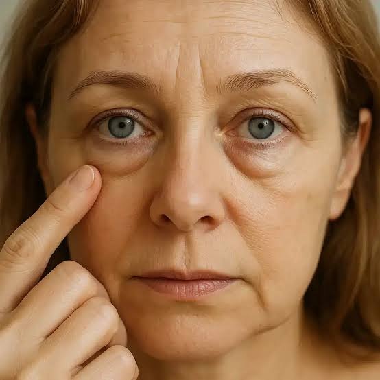
What Causes Eyelid Bags?
Under-eye bags are often described as if they were a single problem, but medically they can represent several very different anatomical changes. One person may have true protrusion of orbital fat. Another may mainly have a deep tear trough and midface hollowing that creates a shadow beneath the lower eyelid. In other patients, morning puffiness, fluid retention, skin laxity, orbicularis muscle prominence or a malar mound may be the dominant feature. These conditions can look similar in a mirror, yet they do not necessarily respond to the same treatment.
This distinction is important because a treatment that improves one anatomical problem can have little effect on another. Removing or repositioning fat may be appropriate when fat prolapse is genuinely responsible for the lower-eyelid bulge, but it does not restore poor skin elasticity. A filler may soften a hollow tear trough in a carefully selected patient, but it is not a universal solution for true fat bags or significant fluid-related swelling. Likewise, treating skin texture cannot correct a deep structural hollow or advanced tissue descent.
A well-known clinical study of 114 consecutive patients seeking consultation for lower-eyelid bags demonstrated exactly this point: most patients had more than one anatomical contributor, and no single category explained the appearance in the majority of cases. The practical message remains highly relevant to modern aesthetic assessment – the correct question is not simply, ‘How do I remove my eye bags?’ It is, ‘What is creating the appearance of my eye bags, and which component should be treated?’
The Six Main Anatomical Causes of Lower-Eyelid Bags
1. Orbital fat prolapse

True orbital fat prolapse is one of the classic causes of a visible lower-eyelid bag. The lower eyelid contains distinct fat compartments. With aging, these compartments can become more visible or project forward, creating a recognizable bulge. The study authors noted that the central fat pad may have a characteristic elongated or ‘cigar-shaped’ contour and that the separate medial, central and lateral fat pockets can sometimes be seen through the skin.
Several mechanisms may make orbital fat look more prominent. Age-related weakening of the orbital septum can allow fat to protrude. At the same time, loss of volume in the cheek and thinning of the tissues around the eye can expose or ‘unveil’ fat that was previously less obvious. This is why an under-eye bag can look more pronounced even when the issue is not simply an increase in the amount of fat.
During examination, true fat prolapse may become more evident in upgaze, and its contour often follows the natural compartmental anatomy of the orbit. Distinguishing this pattern from diffuse fluid is important because fat and fluid behave differently and may require very different management.
2. Eyelid fluid and variable puffiness

Not every puffy lower eyelid is a fat bag. Eyelid fluid can create a smoother, more diffuse swelling that does not respect the separate medial, central and lateral fat compartments. In the source study, fluid-related swelling was described as sometimes having a purplish hue and as changing less with upgaze than orbital fat.
History is especially useful. Fluid-related puffiness may be worse in the morning, after a salty meal or during periods of generalized or local edema. Allergy-related facial swelling may also contribute. A patient who reports that the under-eye fullness changes markedly from day to day may therefore have a different problem from someone with a stable, anatomically defined fat bulge.
This is clinically relevant because simply adding volume beneath a fluid-prone lower eyelid may not address the real cause. A careful consultation should first establish whether the visible fullness is stable fat, variable fluid, a malar mound, a tear-trough shadow or a combination of these findings.
3. Tear-trough depression and midface descent
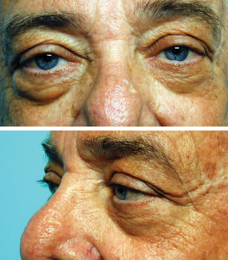
A deep tear trough can make the lower eyelid look heavier even when the apparent ‘bag’ is partly an optical contrast between a bulge above and a hollow below. The tear trough is the depression along the lower orbital rim, extending from the inner under-eye area toward the cheek. It can become more visible as subcutaneous tissue thins, the cheek descends and the transition between the eyelid and midface becomes less smooth.
The source study describes tear-trough depression as an important feature of eyelid and midface aging and notes that it can also be influenced by underlying bony anatomy, including congenital or age-related maxillary hypoplasia. This means some younger patients can have a pronounced tear trough without advanced aging, while older patients may develop a deeper hollow because of both skeletal support and age-related soft-tissue changes.
Tear-trough hollowing is particularly important during treatment planning because reducing a bulge is not always enough. In selected patients, restoring contour at the eyelid-cheek junction may be more important than removing tissue. In others, a combination approach is needed.
4. Loss of skin elasticity, fine lines and sun-related change
The lower eyelid has some of the thinnest skin on the face. As elasticity decreases, the skin can develop fine wrinkles, textural change, crepiness and laxity. Thin or lax skin can also reveal the contours of structures beneath it, including the orbicularis muscle, orbital fat and the tear trough, making pre-existing irregularities look more obvious.
An important principle from the source paper is that traditional fat- or skin-removal surgery is not automatically the best answer for a skin-quality problem. Skin elasticity and surface quality represent their own treatment target. The study’s discussion includes stepwise skin rejuvenation approaches such as skin-care programs, chemical peels and laser resurfacing, while also recognizing that skin treatments cannot compensate for deeper structural problems.
For this reason, a patient whose main concern is fine wrinkling or crepey skin should not be assessed in exactly the same way as a patient with a large fat prolapse or deep tear trough.
5. Orbicularis oculi muscle prominence
The orbicularis oculi is the circular muscle surrounding the eye. In some people, a prominent lower portion of this muscle contributes to a visible roll, particularly during smiling. These lines or rolls can be dynamic, becoming more pronounced with facial expression, although they can coexist with static wrinkles caused by skin aging.
The study authors included orbicularis prominence as a separate anatomical category because it can contribute to the patient’s perception of an under-eye bag even when fat is not the dominant issue. This distinction matters because dynamic muscle prominence is not corrected by the same treatments used for tear-trough hollowing or fat prolapse.
In the paper, conservative botulinum toxin treatment was one of the options used for selected cases of lower-orbicularis prominence. The authors emphasized conservative dosing because excessive weakening of the lower orbicularis can create functional problems. Any treatment in this area therefore requires careful anatomical assessment and appropriate clinical judgment.
6. Triangular malar mound and festoon
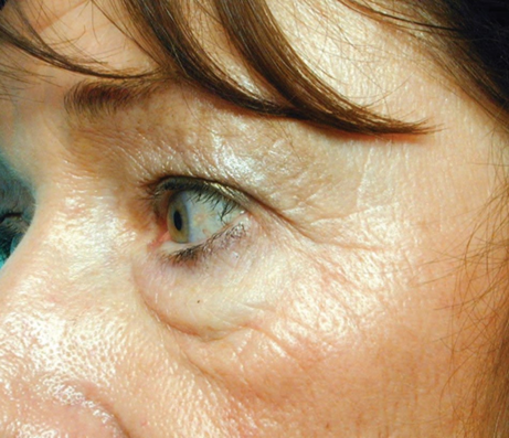
A malar mound or festoon is different from a simple lower-eyelid fat bag. It sits lower, over the upper cheek, and is related to the anatomy of the tissue ‘sponge’ between retaining ligaments around the orbital rim and cheek. The result can be a triangular or crescent-shaped mound that may fluctuate and can become more pronounced when skin elasticity deteriorates.
The source study notes that prominent malar mounds may run in families and may have a variable allergic or fluid-related component. With increasing laxity, a malar mound can progress toward a more obvious festoon. Patients often call this an eye bag because of its location, but the anatomy is different from orbital fat prolapse.
This is one of the reasons the under-eye area can be challenging: a treatment directed at the tear trough or orbital fat may not adequately improve a true festoon. Correct identification of the anatomical level is essential before recommending treatment.
What Did the 114-Patient Study Find?
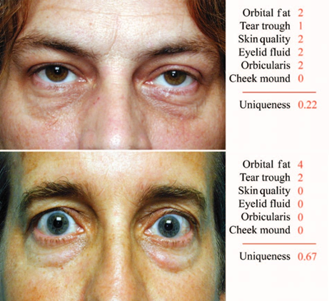
The study evaluated 114 consecutive patients who sought aesthetic consultation for lower-eyelid concerns and had not undergone previous eyelid surgery. There were 67 men and 47 women, with a mean age of 52 years and an age range from 23 to 76 years. Standardized photographs and consultation findings were graded in six anatomical categories.
When the authors calculated the cumulative contribution of each category, the largest contribution came from cheek descent with tear-trough hollowing, followed closely by orbital fat prolapse. Skin laxity and sun damage, eyelid fluid, orbicularis prominence and triangular malar mound also contributed substantially. Importantly, these percentages are not meant to add to 100%, because a single patient could have several problems at the same time.
| Anatomical contributor | Cumulative contribution score |
|---|---|
| Cheek descent / hollow tear trough | 52% |
| Orbital fat prolapse | 48% |
| Loss of skin elasticity / sun damage | 35% |
| Eyelid fluid | 32% |
| Orbicularis prominence | 20% |
| Triangular malar mound / festoon | 13% |
The study’s average ‘uniqueness score’ was approximately 38%, meaning that the most important single feature usually…
The study’s average ‘uniqueness score’ was approximately 38%, meaning that the most important single feature usually accounted for only part of the overall appearance. In practical terms, most patients did not have one isolated cause. The visible bag was commonly the combined result of several anatomical changes.
Why Age Changes the Appearance of Under-Eye Bags
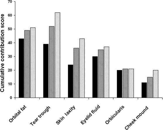
Age does not affect every component of the lower eyelid in the same way. In the study, tear-trough depression and loss of skin elasticity increased particularly with age, and triangular malar mound or festoon was significantly more common in patients older than 50 years. These observations fit the broader concept of facial aging: changes occur in skin, fat, muscle, ligaments, bone and midface support at the same time.
This explains why an older patient may have a combination of a deeper tear trough, more visible orbital fat, thinner skin and cheek descent, while a younger patient may primarily have inherited anatomy, skeletal shape, a naturally deep tear trough or dynamic orbicularis prominence. Age alone does not define the treatment, but it changes the probability that several contributors will coexist.
Are Eye Bags Different in Men and Women?
In this specific study, orbital fat prolapse and tear-trough deformity were somewhat more prominent in men than in women. The average scores for fat prolapse and tear-trough deformity were higher in male participants, with statistically significant differences. This does not mean that men universally develop worse eye bags, but it shows that sex-related anatomical patterns may influence the appearance in a clinical population. Individual assessment remains more important than gender-based assumptions.
How to Tell the Difference: Fat Bag, Tear Trough, Fluid or Festoon?
Patients often use the same phrase – ‘eye bags’ – for several different appearances. A clinical examination looks beyond the label and asks how the contour behaves, where it is located and whether it changes with facial position, expression or time of day.
A compartmentalized bulge that becomes more obvious in upgaze may suggest orbital fat. A diffuse swelling that is worse in the morning or after salty food and does not follow fat compartments may suggest fluid. A fixed depression along the lower orbital rim points toward a tear trough. A roll that becomes more visible with smiling may reflect orbicularis prominence. A mound lower on the cheek, particularly if variable or associated with laxity, may represent a malar mound or festoon.
These distinctions are not always obvious without examination, and mixed patterns are common. A patient can simultaneously have fat prolapse, a tear trough and thin skin. That combination may create a much stronger ‘tired’ appearance than any one feature alone.
Why a One-Size-Fits-All Eye-Bag Treatment Can Fail
The most important clinical lesson from the study is that lower-eyelid rejuvenation should be diagnosis-led rather than procedure-led. Starting with a favorite treatment and then trying to make every patient fit that treatment is the opposite of an anatomical approach.
For example, removing fat in a patient whose primary issue is a hollow tear trough can worsen the transition between the eyelid and cheek. Treating skin texture alone will not correct significant fat prolapse or midface descent. Adding filler beneath an eye that is already prone to fluid-related swelling may not address the underlying problem. And weakening the orbicularis muscle does not treat a fixed fat compartment or festoon.
A good plan therefore identifies the dominant contributors, decides which of them actually need treatment and explains which components may remain even after a successful procedure. This is particularly important in the lower eyelid, where small contour changes can be visually significant.
Treatment Options Should Match the Anatomy
1. When orbital fat is the main problem

For patients with substantial true orbital fat prolapse, the source paper describes lower-eyelid blepharoplasty and fat repositioning as surgical options. Traditional transconjunctival blepharoplasty with fat removal may still have a role when the fat itself is clearly prominent. However, modern lower-eyelid planning often aims to preserve or reposition volume when appropriate rather than treating every bag by simply removing fat.
Fat repositioning may be considered when there is adequate orbital fat together with a significant tear-trough depression, because moving existing fat can help smooth the transition across the orbital rim. Whether surgery is appropriate depends on anatomy, skin quality, support, symptoms, medical history and the patient’s goals.
2. When tear-trough hollowing is the main problem
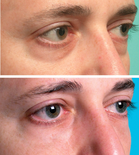
When the dominant feature is a hollow tear trough rather than a large protruding fat bag, volume restoration can become more relevant. The source article discusses fat injection and cross-linked hyaluronic acid filler as options for periorbital volume augmentation in selected cases.
This does not mean that every tear trough should be filled. Under-eye filler requires careful patient selection because the area is anatomically delicate and because edema, skin quality, fat prolapse and the shape of the eyelid-cheek junction can affect the result. The objective is not simply to ‘fill the line’ but to improve the contour while respecting the surrounding anatomy.
3. When skin quality is the main problem
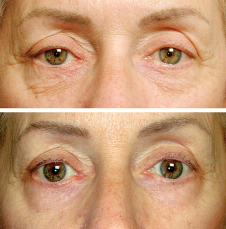
For fine wrinkling, sun-related changes and reduced elasticity, the source study discusses skin-care programs, chemical peels and laser resurfacing as skin-focused approaches. The authors also make an important limitation clear: skin rejuvenation cannot correct deep structural problems.
If a patient has severe skin redundancy or advanced festoon formation, a purely non-surgical skin treatment may not be enough. Conversely, removing skin without addressing poor skin quality does not necessarily restore elasticity or texture.
4. When orbicularis prominence is the main problem
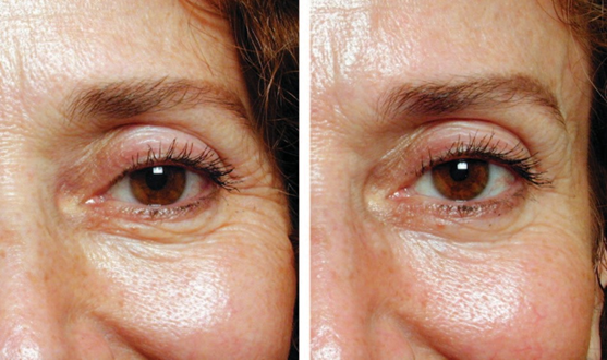
For selected dynamic lower-eyelid orbicularis prominence, the study includes conservative botulinum toxin treatment. This is a highly technique-sensitive area. Excessive weakening can affect lower-eyelid function, which is why conservative, individualized dosing and appropriate clinical expertise are essential.
5. When fluid or festoons are the main problem
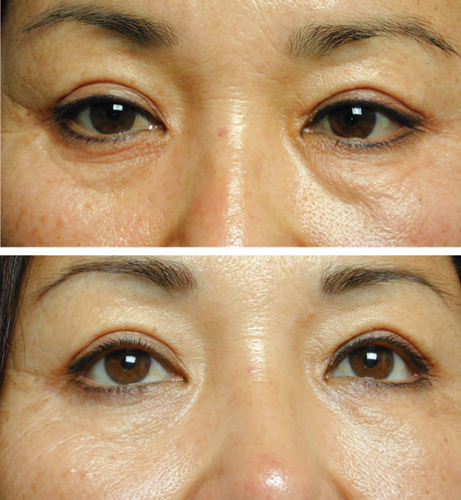
Fluid-dominant lower-eyelid swelling and festoons require a different diagnostic mindset. The source paper describes a radiofrequency technique used for the eyelid fluid sponge in its historical treatment series, but the broader lesson is more important than any single procedure: a fluid-prone or festoon pattern should not be assumed to behave like a tear trough or ordinary fat bag.
Because swelling can be influenced by local or systemic factors, a clinician may also need to consider whether there are non-aesthetic causes or triggers that require further medical evaluation. Persistent or unexplained swelling should not automatically be treated as a cosmetic contour problem.
Non-Surgical Treatment vs. Surgery : What Is the Difference?
Non-surgical treatments can be very useful when the target is mild to moderate contour irregularity, selected tear-trough hollowing, dynamic muscle prominence, skin quality or certain forms of tissue laxity. Their main advantage is that they can address specific components without the recovery associated with surgery. Their limitation is equally important: they cannot reproduce every structural change that surgery can achieve.
Surgery becomes more relevant when there is substantial fat prolapse, significant tissue descent, marked skin redundancy or an anatomical problem that cannot be predictably corrected with a minimally invasive option. The source study’s regression analysis found that recommendations for lower blepharoplasty were influenced by the extent of fat prolapse and skin laxity, while tear-trough deformity also influenced the recommendation for other surgical procedures.
The best approach is therefore not ‘non-surgical is always better’ or ‘surgery is always more effective.’ The correct approach depends on the component being treated and the degree of anatomical change.
Who May Not Be an Ideal Candidate for Under-Eye Filler?
A patient can have a visible tear trough and still be a poor candidate for filler if the surrounding anatomy is unfavorable. A prominent true fat bag, significant fluid retention, advanced festoon, marked skin laxity or a complex eyelid-cheek contour may limit what filler can realistically achieve.
This is one of the practical implications of understanding the six anatomical categories. If a hollow is only one small part of a larger pattern, filling it alone may produce an incomplete result. A consultation should therefore assess both the depression and the tissues above and below it rather than focusing on a single line in isolation.
What Should Be Assessed During an Under-Eye Consultation ?
A structured lower-eyelid assessment should evaluate multiple components rather than looking only at the visible bag. Important points include :
- Whether the fullness is stable or changes during the day.
- Whether the contour is compartmentalized like orbital fat or diffuse like fluid.
- The depth and shape of the tear trough and eyelid-cheek junction.
- Midface volume and cheek descent.
- Skin thickness, laxity, texture, sun damage and fine wrinkling.
- Orbicularis activity at rest and with smiling.
- Presence of a malar mound or festoon below the lower eyelid.
- Previous surgery, filler, energy-based treatments or other procedures around the eyes.
- Relevant medical history, allergy-related swelling and any persistent or unexplained edema.
- The patient’s actual goal: less puffiness, a smoother contour, improved skin quality, fewer lines or a more rested appearance.
Why the Eyelid-Cheek Junction Matters So Much
Many patients perceive ‘bags’ because the transition from the lower eyelid to the cheek has become uneven. A small convexity of fat can look much larger when it sits directly above a deep tear trough. Conversely, improving a hollow can sometimes make the entire lower eyelid appear smoother even though the orbital fat itself has not disappeared.
This is why lower-eyelid assessment should include the midface. The study’s highest cumulative contribution score was actually the combination of cheek descent and hollow tear trough, slightly higher than orbital fat prolapse. The lower eyelid cannot always be treated as an isolated structure.
Can Eye Bags Be Genetic ?
Yes, the appearance of the lower eyelid can be strongly influenced by inherited anatomy. The source article notes that tear-trough depression can relate to underlying bony structure and that prominent triangular malar mounds may run in families. A younger person can therefore have noticeable lower-eyelid hollowness or mounding without the same degree of age-related skin change seen in an older patient.
Yes, the appearance of the lower eyelid can be strongly influenced by inherited anatomy. The source article notes that tear-trough depression can relate to underlying bony structure and that prominent triangular malar mounds may run in families. A younger person can therefore have noticeable lower-eyelid hollowness or mounding without the same degree of age-related skin change seen in an older patient.
Can Salt, Sleep or Allergies Make Under-Eye Bags Worse ?
They can influence fluid-related puffiness. In the source study, eyelid fluid was described as being worse after a salty meal or in the morning, and local facial allergy was mentioned as one setting in which edema may accumulate in the eyelid tissues. This kind of fluctuation is clinically useful because it suggests that the apparent bag may not be composed entirely of fat.
However, lifestyle factors do not explain every under-eye bag. A fixed fat prolapse, deep tear trough, skin laxity or inherited malar mound may remain visible even when sleep, hydration and diet are optimized.
A More Precise Way to Think About “Tired Eyes”
The phrase ‘tired eyes’ is a visual description, not an anatomical diagnosis. Darkness may come from a hollow casting a shadow. Puffiness may be fat or fluid. Wrinkles may be caused by skin laxity or muscle activity. A lower-cheek mound may be a festoon. Several of these can occur together, creating a tired appearance even in a healthy, well-rested person.
A precise diagnosis helps set realistic expectations. If the appearance is mostly a shadow created by a tear trough, reducing swelling will not fully solve it. If the issue is a true fat bulge, filling the surrounding area may camouflage the contour but does not remove the fat. If the problem is skin quality, structural treatment alone may leave crepiness and fine lines unchanged.
