
Under-Eye Filler in Abu Dhabi : A Complete Evidence-Based Guide to Tear Trough and Periorbital HA Treatment
The under-eye region is one of the most technically demanding areas in non-surgical facial rejuvenation. A patient may describe tired eyes, dark circles, hollowness, a visible lid-cheek junction, crow’s-feet lines or loss of support around the outer eye, yet these concerns do not all arise from the same anatomy. Hyaluronic acid filler can be highly effective when the dominant problem is a treatable contour deficit, but the choice of product, injection plane, amount and patient selection are especially important in this thin and anatomically sensitive region.
The 2025 EYELIGHT study provides useful real-world evidence because it did not force clinicians to use one rigid technique. Instead, practitioners treated patients in routine clinical practice and selected indication, volume, depth and technique according to their normal judgment. This makes the data particularly relevant to how periorbital filler is actually used outside a tightly controlled trial, while also creating limitations because treatment was heterogeneous.
The study focused on TEOSYAL PureSense Redensity 2, a hyaluronic acid filler developed for the under-eye region. Among 136 participants, investigators used the product not only in the tear trough but also in the palpebromalar groove, outer canthus, crow’s-feet region and brow. The results support meaningful aesthetic improvement in selected patients, high satisfaction and a generally favorable short- to medium-term safety profile, with improvement still perceived in some patients at 12 months.
Why the Periorbital Region Requires Special Product Selection
The under-eye is not an ideal place for a generic volumizing filler. The skin is thin, the transition from eyelid to cheek is highly visible, and the region is prone to swelling. The source article explains that HA fillers differ substantially in strength, cohesivity, malleability, stretch, concentration, molecular weight and degree of cross-linking. These rheological properties influence how a gel integrates into tissue and how it behaves after injection.
The R2 product evaluated in EYELIGHT is described as having relatively low strength and rigidity, low resistance to compression, high malleability and spreadability, and a low HA concentration. It combines cross-linked and non-cross-linked high-molecular-weight HA. The authors state that this design minimizes water absorption and therefore aims to reduce product-associated swelling, an especially important consideration in the under-eye.
This study does not prove that the same results apply to every HA filler. A major practical lesson is that evidence from one periorbital product should not automatically be generalized to products with different rheology or water-binding behavior.
What Is a Tear Trough?
The tear trough is the hollow that extends from the inner lower eyelid toward the upper cheek. When this depression becomes more visible, it can create a shadow that makes the face look tired even when the skin itself is not heavily pigmented. In appropriate patients, a small amount of carefully selected HA filler can soften this transition and reduce the contrast between the lower eyelid and cheek.
A tear trough is not the same as lower-eyelid puffiness, malar edema or prominent orbital fat. Filler can camouflage some contour differences, but it may be unsuitable when puffiness or fluid retention is the dominant problem. This distinction is central to avoiding overfilling and prolonged swelling.
The EYELIGHT registry studied several periorbital indications separately. Tear trough was the most common initial indication, representing 35.3% of initial periorbital R2 treatments. Palpebromalar groove represented 24.2%, outer canthus 17.9%, crow’s feet 15.1% and brow 7.5%.

What Was the EYELIGHT Study?
EYELIGHT was a multicenter prospective observational study conducted in Europe. Patients were treated between May 2020 and February 2021 across two centers in the United Kingdom and one in France, with follow-up data collected until July 2022. The study enrolled adults seeking non-surgical aesthetic treatment who met the eligibility criteria.
The study design intentionally reflected routine clinical practice. Every enrolled subject received at least one R2 injection. Treatment of one or multiple indications was allowed, and clinicians could choose volume, depth and injection technique. Follow-up visits were planned at 3, 6, 9 and 12 months, although visits were optional because of the observational design.
The primary efficacy endpoint was the proportion of treatments rated ‘improved’ or ‘much improved’ on the Global Aesthetic Improvement Scale (GAIS) at approximately three months, as judged by both the investigator and the subject. Safety monitoring recorded common treatment responses such as bruising, swelling, redness and lumps, as well as adverse events.
Who Was Included in the Study?
A total of 136 subjects were enrolled and 129 completed the study. The population was predominantly female: 122 participants, or 89.7%, were women and 14, or 10.3%, were men. Mean age was 48.4 years, with a range from 23 to 77 years. Most participants had Fitzpatrick skin type II or III.
The study excluded people with several conditions or recent treatments that could complicate interpretation or safety. Examples included filler injection in the same area within the preceding six months, active local skin disease or infection, known hypersensitivity to lidocaine/local anesthetics or HA, certain autoimmune, cardiac, hepatic and neurologic conditions, planned peeling or laser/ultrasound procedures soon after injection, pregnancy and breastfeeding.
What this means for website claimsThe results describe a selected study population. They should not be interpreted as proof that every person with an under-eye concern is suitable for filler. Medical history, previous filler, edema tendency, skin quality and local anatomy can materially change treatment suitability.
How Was the Product Used in Real Practice?
Across the overall study, 958 injections were performed at the initial visit, and 451 were performed with R2 alone. For the periorbital efficacy analysis, 252 initial R2-only injections treated one of five periorbital indications. Most tear trough, palpebromalar groove and outer-canthus treatments were placed deeply toward the supraperiosteal plane, whereas crow’s-feet and brow treatments were more often superficial.
The average volume of R2 used for one indication was 0.32 ± 0.25 mL, with a reported range from 0.1 to 1.0 mL. For tear trough treatment, the mean total volume for both sides was 0.55 ± 0.37 mL. These are descriptive study averages rather than recommended doses for every patient.
Most periorbital treatments were performed without pre-procedure anesthesia. Approximately one-third of tear-trough injections used topical anesthetic. Only a small proportion of tear trough and palpebromalar injections used a cannula or an alternative needle, reflecting the operators’ routine preferences.
Only 24 of the 136 subjects required a touch-up and 12 required retreatment during the study. That finding is relevant to durability, although it is important to remember that follow-up visits were optional and subjects could have other aesthetic indications treated during the registry.
How Effective Was Under-Eye and Periorbital HA Filler at 3 Months?
The primary analysis included 283 R2 treatments with available month-3 GAIS data. Across all indications, 214 treatments, or 75.6%, were rated ‘improved’ or ‘much improved’ by both investigator and subject. The 95% confidence interval was 70.6% to 80.6%, meaning the study met its predefined primary effectiveness threshold.
A sensitivity analysis broadened the acceptable time window and used directly post-injection scores when later data were missing. In that analysis, 355 of 434 treatments, or 81.8%, were rated improved or much improved by both investigator and subject.
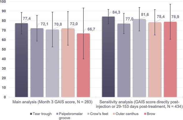
When analyzed by periorbital indication, the main month-3 analysis showed improvement rates above 70% for tear trough, palpebromalar groove, crow’s feet and outer canthus. Brow was 66.7% in the smaller main-analysis sample but reached 78.9% in the sensitivity analysis.
| Periorbital indication | Main month-3 GAIS: improved/much improved | Sensitivity analysis |
|---|---|---|
| Tear trough | 41/53 (77.4%) | 75/89 (84.3%) |
| Palpebromalar groove | 31/43 (72.1%) | 47/61 (77.0%) |
| Crow’s feet | 17/24 (70.8%) | 31/38 (81.6%) |
| Outer canthus | 18/25 (72.0%) | 29/37 (78.4%) |
| Brow | 8/12 (66.7%) | 15/19 (78.9%) |
How Long Did Improvement Last?
The highest improvement rates were seen immediately after injection, which is consistent with the immediate volumizing effect of HA. Investigator ratings remained higher than subject ratings at every later timepoint.
According to the investigators, at least 82% of subjects were still considered improved at month 9 and at least 60% at month 12 across the reported indications. Subject ratings were more conservative: improvement remained evident in approximately 56% to 66% at month 9 and 48% to 51% at month 12, depending on the area.
The palpebromalar groove had the highest month-12 improvement among the reported indications, rated improved by 85% of investigators and 51% of subjects. Brow data were not available at month 12.
The difference between investigator and patient ratings is clinically important. The authors suggest that patients may have higher expectations and may also become accustomed to their post-treatment appearance, whereas investigators can compare standardized photographs against baseline.

Patient Satisfaction and the ‘Natural-Looking’ Result
At month 3, satisfaction was high for several indications. Eighty-five percent of subjects were satisfied or very satisfied with tear trough treatment, 83% with the palpebromalar groove and 92% with the outer canthus. The source text reports 69% for the brow; satisfaction data for crow’s feet were not presented in the same summary.
Recommendation rates were also high: 93% of tear trough patients, 83% of palpebromalar groove patients and 95% of outer-canthus patients were somewhat or very likely to recommend the treatment.
Most patients selected ‘natural looking’ as the word that best described the outcome: 71% for tear trough, 72% for palpebromalar groove, 74% for outer canthus and 63% for brow. This is useful because under-eye rejuvenation is an area where a subtle transition is often more aesthetically desirable than obvious volume.
Almost all investigators, 99%, reported being satisfied with the product in the periorbital area, and 100% described it as easy to inject. These investigator impressions are relevant but should be interpreted in context because the study was industry funded.
Before-and-After Images : How Should They Be Interpreted?
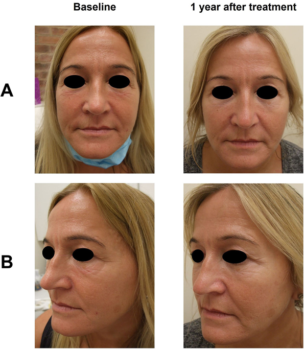
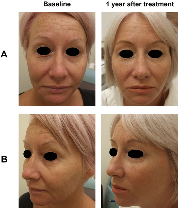
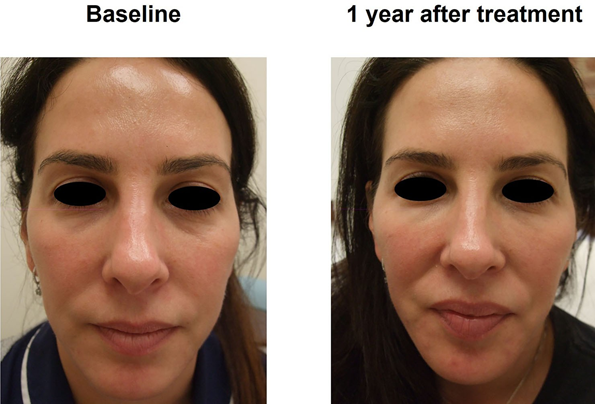
The source article provides representative images at baseline and one year after treatment. These photographs are helpful for visualizing the type of change reported in the registry, but they are not isolated tear-trough filler experiments. The figure captions state that the subjects also received treatment in multiple additional facial areas.
Therefore, the photographs should not be used to claim that every visible facial difference is caused by R2 in the periorbital region. The most scientifically accurate interpretation is that they illustrate real-world patients who received periorbital R2 as part of broader individualized aesthetic treatment.
What Was the Safety Profile in the Study?
Safety is particularly important around the eyes because even mild swelling or contour irregularity can be visually obvious. In EYELIGHT, the common treatment responses reported immediately after injection were all graded mild or moderate and resolved within one month.
For tear trough injections, bruising occurred after 17 of 116 injections (14.7%), swelling after 9 (7.8%), redness after 6 (5.2%) and lumps or bumps after 1 (0.9%). The palpebromalar groove had bruising in 9.0% and swelling in 2.6%. In the crow’s-feet region, lumps or bumps occurred in 14.6%, bruising in 10.4%, moderate redness in 2.1% and swelling in 2.1%. Outer-canthus treatments had bruising in 11.3%, and brow treatments had swelling in 20.0% and bruising in 10.0%.
Only four adverse events were reported among the 136 subjects, and two were considered device related. Only one device-related adverse event followed a periorbital injection: moderate skin edema after brow treatment. It resolved without sequelae after 28 days.
The authors also emphasize an important broader point: delayed edema after HA filler can occur and may persist well beyond the immediate treatment period in rare cases. The favorable EYELIGHT data should therefore not be interpreted as evidence that delayed complications are impossible.
| Area | Bruising | Swelling | Other notable immediate CTRs |
|---|---|---|---|
| Tear trough | 14.7% | 7.8% | Redness 5.2%; lumps/bumps 0.9% |
| Palpebromalar groove | 9.0% | 2.6% | Moderate redness 1.3% |
| Crow’s feet | 10.4% | 2.1% | Lumps/bumps 14.6%; moderate redness 2.1% |
| Outer canthus | 11.3% | 1.9% | Redness 1.9%; lumps/bumps 1.9% |
| Brow | 10.0% | 20.0% | Lumps/bumps 5.0% |
How Painful Was Treatment?
Mean injection pain on a 100-mm visual analog scale was generally in the 20-30 mm range, which the authors interpreted as mild. Mean pain scores during injection were 20.3 mm for tear trough, 26.1 mm for palpebromalar groove, 24.8 mm for crow’s feet, 26.7 mm for outer canthus and 22.8 mm for brow.
By the end of the visit, mean pain scores were close to zero in all areas. A few individuals reported higher pain during tear-trough injection, including scores up to 80 mm, but this was short lived.
Safety wording suitable for a medical websiteA source-grounded statement is: ‘In the EYELIGHT registry, immediate treatment reactions were mild or moderate and resolved within one month; no severe adverse events were reported, and one device-related periorbital adverse event—moderate brow edema—resolved after 28 days.’ This should not be rewritten as ‘risk-free.’
Who May Be a Good Candidate for Under-Eye HA Filler?
The source study was not designed to create a universal candidacy algorithm, but its design and exclusion criteria reinforce several practical principles. A patient is more likely to benefit when the visible concern is an appropriate contour deficit rather than primarily fluid retention, severe skin excess or another problem that filler cannot solve.
- A visible tear trough or lid-cheek contour depression that can be improved with small-volume structural correction.
- Realistic expectations for subtle rather than exaggerated volume change.
- No active infection or inflammatory skin condition near the treatment area.
- No recent filler in the same area that would make anatomy and residual product difficult to interpret.
- No contraindication identified from medical history, medication history or allergy history.
- An anatomy in which the clinician believes the expected aesthetic benefit outweighs the risk of swelling or contour irregularity.
The source cannot support a claim that every patient with dark circles is suitable. Patients with puffiness, malar edema, prominent lower-eyelid fat or a primarily pigment-based problem may need a different strategy.
Why Overfilling the Under-Eye Is a Problem
HA works by occupying space and interacting with water. In a region where the skin is thin and the lymphatic environment is delicate, excessive volume or an unsuitable gel can produce visible swelling, irregularity or a heavy appearance. The source article specifically emphasizes product characteristics that aim to minimize water absorption in the under-eye.
This reinforces a conservative treatment philosophy. The EYELIGHT tear-trough mean volume was approximately 0.55 mL total for both sides, but this is not a dosing rule. Some patients need less, some may not be suitable for direct tear-trough injection, and others may benefit from treatment of neighboring structural support rather than placing more product directly into the hollow.
A good result in the under-eye is not measured by how much filler is injected. It is measured by whether the eyelid-cheek transition looks smoother without producing puffiness, visible product or an unnatural contour.
Under-Eye Filler vs Midface Support
The source study allowed clinicians to treat multiple facial areas, and the representative subjects often received treatment beyond the periorbital region. This reflects real-world practice: an apparent tear trough can be influenced by cheek volume, the palpebromalar groove and the overall midface contour.
The article does not provide a controlled comparison of direct tear-trough filler versus midface-first strategies. Therefore, it cannot prove which sequence is best. It does, however, support an individualized whole-face approach rather than treating every under-eye hollow as an isolated line.
For patients, this means an assessment should explain whether the perceived tired appearance comes from the tear trough itself, from adjacent support loss, from skin quality, or from a combination.
What the Study Does and Does Not Prove
| The study supports | The study does not establish |
|---|---|
| Real-world aesthetic improvement with R2 in five periorbital indications | That all HA fillers behave the same way in the under-eye |
| High 3-month GAIS improvement in selected patients | That every patient with dark circles is a filler candidate |
| Improvement perceived in a proportion of patients through 12 months | A guaranteed 12-month duration for every individual |
| Generally mild or moderate common treatment responses in this registry | That serious or delayed HA-filler complications are impossible |
| High satisfaction and frequent ‘natural-looking’ patient descriptions | That the representative photographs reflect R2 alone, because subjects also received other treatments |
| Routine clinical use beyond the tear trough in this dataset | Regulatory approval of the product for every periorbital indication in every jurisdiction |
Study Limitations and Potential Bias
EYELIGHT is valuable because it reflects routine clinical practice, but observational evidence is inherently less controlled than randomized evidence. Clinicians selected treatment indications, volume, depth and technique, so the data include real-world variability that can improve generalizability while making direct comparisons more difficult.
Follow-up visits were optional, which created missing data at later timepoints. The authors addressed missingness using sensitivity analyses, but missing data can still influence interpretation. Subject and investigator ratings also differed, with investigators generally reporting greater improvement.
Industry relationships are another important context. The study was funded by Teoxane S.A. Two authors were employees of Teoxane, and the clinical investigators received research grants for study participation. These disclosures do not invalidate the data, but they are relevant when interpreting favorable product-specific conclusions.
Finally, the study evaluated one specific HA filler. Its results should not be automatically transferred to other under-eye fillers with different HA concentration, cross-linking, rheology or tissue integration.
A Practical Consultation Framework for Tear Trough Filler
- Define the patient’s primary concern: hollowing, shadowing, puffiness, skin quality, pigment or a combination.
- Examine lower-eyelid and midface anatomy rather than treating the tear trough as an isolated line.
- Assess previous filler history and the possibility of residual product.
- Assess edema tendency and whether swelling is already present before treatment.
- Discuss whether direct tear-trough correction, adjacent support treatment, another modality or no filler is the safer choice.
- Use conservative volume and an appropriate periorbital product when filler is selected.
- Document baseline photographs so improvement and delayed change can be judged against the original anatomy.
- Explain both immediate reactions and the possibility of delayed swelling or contour issues.
- Set realistic expectations: improvement is common in well-selected patients, but perfect correction is not guaranteed.
