
Lower Eyelid Blepharoplasty in Abu Dhabi: A Complete Evidence-Based Guide
Lower eyelid bags are often described as a simple problem of “excess fat,” but modern eyelid surgery is much more nuanced. The appearance of the lower eyelid is created by the interaction of skin quality, orbital fat, the orbital septum, the orbicularis muscle, the tear trough, the canthal support system and the contour of the midface. A successful lower eyelid rejuvenation plan therefore begins with diagnosis rather than with a single operation.
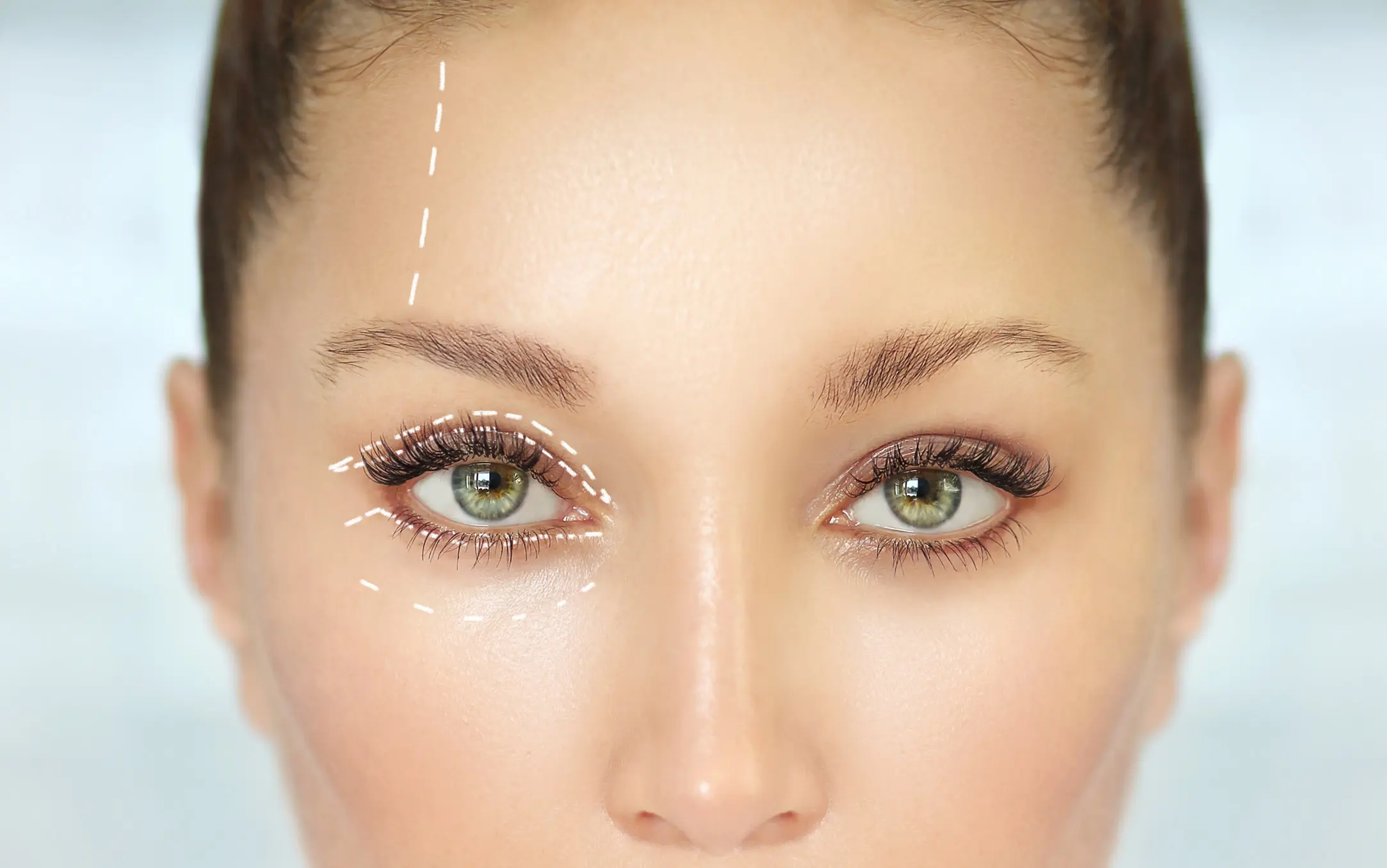
Lower eyelid blepharoplasty is a surgical procedure designed to improve selected age-related or structural changes of the lower eyelid. Depending on the patient, the goal may be to reduce prominent eye bags, remove genuinely redundant skin, reposition orbital fat into a hollow tear trough, improve the lower lid-cheek transition, support a lax lateral canthus or combine several of these objectives in one individualized plan.
The evidence reviewed in the source article reflects an important change in aesthetic surgery: the historical philosophy of simply removing skin and fat has gradually shifted toward tissue preservation, conservative excision and volume redistribution. Complete removal of the lower eyelid fat pads can leave some patients looking hollow or skeletonized, while thoughtful preservation and repositioning may create a smoother transition from the lower eyelid to the cheek.
For patients researching lower eyelid blepharoplasty in Abu Dhabi, this distinction matters. Two people can both complain of “bags under the eyes” while having entirely different anatomy. One may have isolated orbital fat prolapse with good skin tone. Another may have excess skin, laxity of the lower eyelid, a prominent tear trough, flat malar projection or a combination of these features. The most appropriate treatment—and whether surgery is even the right treatment—depends on the pattern identified during clinical assessment.
What Is Lower Eyelid Blepharoplasty?
Lower eyelid blepharoplasty is an umbrella term rather than one fixed surgical maneuver. It includes a family of procedures that address the lower eyelid and the lower eyelid-cheek junction. Traditional operations focused mainly on excision of protruding fat and loose skin. Contemporary approaches may still remove carefully selected excess tissue, but often combine that with fat preservation, fat transposition, canthal support, skin resurfacing or midface procedures when the anatomy requires them.
The two principal surgical routes are the transconjunctival approach and the transcutaneous approach. In a transconjunctival procedure, the lower eyelid fat compartments are accessed from the inside of the eyelid. This is particularly relevant in younger patients or in patients with prominent fat prolapse but minimal excess skin. In a transcutaneous procedure, the lower eyelid is approached through the skin, generally through a subciliary incision, allowing the surgeon to directly treat redundant skin and combine the operation with other lower-lid support procedures when indicated.
The review emphasizes that the transconjunctival approach is frequently preferred because it can avoid an external skin incision and may carry less risk of visible scarring or certain forms of lower-lid malposition in properly selected patients. It is not, however, a substitute for the transcutaneous approach when meaningful skin excess must be corrected. The operation has to fit the anatomy rather than the other way around.
Why Do Eye Bags and Lower Eyelid Aging Develop?
The aging lower eyelid is a multi-layered problem. Changes can occur in the skin, the orbital septum, the canthal tendons, the orbicularis oculi muscle and the deeper fat compartments. The result can be a mixture of puffiness, wrinkles, tear trough hollowing, lengthening of the lower eyelid, festoons, malar mounds and an increasingly visible orbital rim.
- Skin laxity and dermatochalasis can create creasing, redundancy and a tired lower-eyelid appearance.
- Weakening or stretching of the orbital septum can allow orbital fat to become more prominent.
- Orbital fat prolapse can create the classic bulging appearance of lower eyelid bags, also called steatoblepharon.
- Tear trough hollowing may become more visible because of volume loss, descent of tissues or underdevelopment of the infraorbital-malar complex.
- Canthal tendon laxity and reduced lower-lid tone may make the eyelid less stable and increase the risk of postoperative malposition if not recognized.
- Malar deflation or a relatively flat cheek can exaggerate the step-off between the lower eyelid and the midface.
- Festoons and malar mounds represent a different contour problem and may not respond to simple fat removal alone.
One of the most important aesthetic concepts is the lower lid-cheek junction. A youthful lower eyelid tends to blend smoothly into the cheek. With aging, a convex bulge of orbital fat may sit above a concavity at the infraorbital rim and a second convexity over the malar mound. The review describes this as a “double convex” pattern. Modern lower eyelid rejuvenation tries not merely to flatten the upper bulge but to restore a more continuous contour across the entire lower eyelid-midface unit.
Lower Eyelid Anatomy : Why Precise Assessment Matters
Lower eyelid surgery takes place in a compact region containing delicate structures that influence eyelid support, eye movement and facial expression. The review divides the lower eyelid into three lamellae. The anterior lamella contains the skin and orbicularis oculi muscle. The middle lamella contains the orbital septum. The posterior lamella contains the tarsal plate, eyelid retractors and palpebral conjunctiva.
The orbital septum is a thin fibrous diaphragm separating orbital contents from the superficial tissues of the face. The lower eyelid retractors, often described in relation to the capsulopalpebral fascia, insert near the lower border of the tarsus. Laterally, the orbital retaining ligament connects the orbicularis region to the orbital rim and contributes to the contour of the eyelid-cheek transition.
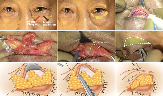
The review highlights three retroseptal lower eyelid fat pads: medial, central and lateral. The inferior oblique muscle lies between the medial and central fat compartments, which is clinically important because injury to this muscle may cause diplopia. The central and lateral fat pads are separated by the arcuate expansion. This anatomy explains why “eye bags” are not one uniform mass and why aggressive or poorly directed fat removal can create contour problems.
Another relevant concept is the surgical “critical zone” described in the source review. It is located inferolateral to the lateral canthus and contains terminal branches that supply the pretarsal and preseptal orbicularis. Injury in this region can contribute to weakness or postoperative ectropion. For patients, the practical lesson is simple: lower eyelid blepharoplasty requires detailed knowledge of both aesthetic anatomy and eyelid function.

Tear Trough Deformity and the Importance of Volume
Tear trough deformity is one of the reasons modern lower eyelid surgery has moved away from indiscriminate fat removal. The tear trough is the hollow that may run from the inner lower eyelid toward the upper cheek. It can become more visible because of volume loss, descent of facial tissues, skeletal anatomy or a combination of these factors.
If a patient has prominent fat above a hollow orbital rim, removing all visible fat may reduce the bulge but deepen the hollow. The eye can then look more sunken or skeletonized. The source review discusses the historical evolution from simple fat excision toward fat preservation and transposition. Instead of discarding all prolapsed fat, selected fat can be mobilized and repositioned over the infraorbital rim to soften the lower lid-cheek junction.
The concept is not that every patient needs fat repositioning. Some patients genuinely benefit from conservative fat excision, some from relocation, and others from a combination. The key is to treat the convexity and the hollow as parts of the same three-dimensional contour problem.
Who May Be a Candidate for Lower Eyelid Blepharoplasty?
The source review lists several common indications, including lower eyelid dermatochalasis and rhytidosis, relative steatoblepharon, a pronounced nasojugal groove, infraorbital or malar deflation, malar mounds or festoons, and lower eyelid asymmetry. However, the presence of one of these features does not automatically mean that surgery is indicated.
Candidate selection should consider the amount of skin excess, the degree of orbital fat herniation, the depth of infraorbital hollowing, the laxity of the lateral canthus, overall lower-lid tone and the vector relationship between the lower eyelid and cheek. In general, the review supports a transconjunctival route for younger patients with fat herniation and minimal skin excess, whereas a transcutaneous route becomes more relevant when significant excess skin needs to be removed.
Aesthetic diagnosis comes before techniqueA patient with true orbital fat prolapse, a patient with tear trough hollowing and a patient with skin laxity can all use the phrase “under-eye bags,” but the correct treatment may be completely different.
Preoperative Evaluation: What Should Be Assessed?
A careful preoperative evaluation is one of the strongest recurring themes in the review. Lower eyelid surgery is not simply a cosmetic skin procedure; it involves structures that protect the eye and maintain eyelid position. The assessment should therefore include both aesthetic and ophthalmic considerations.
- Medical history and previous eye or eyelid surgery.
- Lower eyelid position and the amount of inferior scleral show.
- Periocular skin quality, true skin redundancy and rhytides.
- Prominence of the medial, central and lateral orbital fat compartments.
- Tear trough depth and infraorbital hollowing.
- Dry-eye symptoms and ocular-surface considerations.
- Horizontal lower-lid laxity and canthal tendon laxity.
- Cheek projection and malar support.
- Malar mounds or festoons.
- Negative-vector anatomy and the relationship between the orbital rim and the lower eyelid.
- Baseline asymmetry and standardized preoperative photographs.
This is also why a photo alone cannot determine the correct operation. Dynamic examination, lid tone, eye-surface symptoms and skeletal support may change the plan. A conservative surgical plan that respects the patient’s support structures may be more important than trying to remove every visible irregularity in a single step.
Transconjunctival vs Transcutaneous Lower Eyelid Blepharoplasty
| Feature | Transconjunctival | Transcutaneous | Main advantage | Main limitation |
|---|---|---|---|---|
| Incision | Inside lower eyelid conjunctiva | Below lash line / skin approach | No external skin incision with transconjunctival approach | Transconjunctival approach cannot directly remove significant excess skin |
| Best suited to | Prominent fat prolapse with little skin excess | Patients with clinically significant skin redundancy | Approach can be matched to anatomy | Patient selection is essential |
| Fat management | Conservative excision and/or repositioning | Excision and/or pedicled repositioning | Modern philosophy favors preservation when possible | Over-resection can create hollowing |
| Canthal support | May be added when laxity is present | Canthal support may be added | Helps address lower-lid support | Adds complexity and must be individualized |
| Scarring / lid position | Potentially less external scarring and lower ectropion risk in selected patients | Useful when skin excision is required | Each technique solves a different problem | Transcutaneous surgery has greater concern for retraction/scleral show in susceptible patients |
Transconjunctival Lower Eyelid Blepharoplasty
The transconjunctival approach accesses the lower eyelid fat from the inside of the eyelid. Because there is no external skin incision, it has become an important technique for patients who primarily have orbital fat prolapse without meaningful skin redundancy. The review describes a broad shift among oculoplastic surgeons toward this approach, although it remains one tool rather than a universal replacement for transcutaneous surgery.
The main aesthetic advantage is that the surgeon can address prominent lower eyelid fat while preserving the external skin envelope. Depending on the patient, fat may be conservatively excised, repositioned or both. The review notes that many surgeons use the lower eyelid fat as pedicled tissue to fill the infraorbital hollow rather than simply discarding it.
From a patient perspective, the most important issue is not the technical name of the incision but whether the chosen approach matches the actual problem. If the lower eyelid skin is already tight and the dominant issue is fat prolapse, an external skin incision may add unnecessary morbidity. If there is substantial skin excess, however, a transconjunctival-only procedure may leave the skin problem untreated.
Transcutaneous Lower Eyelid Blepharoplasty
The transcutaneous approach is most relevant when the patient has lower eyelid skin redundancy that truly requires excision. It generally provides direct access to the skin, orbicularis and orbital septum, and it can be combined with canthopexy, canthoplasty or midface procedures when additional support or contour correction is required.
The source review describes the “skin pinch” concept as a way to estimate skin laxity in selected cases and emphasizes conservative skin removal. Excessive removal or damage to the orbicularis can destabilize the lower eyelid and contribute to retraction, scleral show or ectropion. This is one reason why modern lower eyelid surgery increasingly favors conservative, support-preserving techniques.
The transcutaneous route therefore has a very important role, particularly in patients with excess skin, but it demands careful planning of both tissue removal and eyelid support. In the review, the possibility of visible scarring and postoperative lower-lid malposition is one of the factors that makes surgeons more selective about using this approach when a transconjunctival option could adequately address the problem.

Fat Excision vs Fat Preservation and Repositioning
Perhaps the most important conceptual evolution described in the review is the move from aggressive fat excision toward fat preservation. In the older model, protruding fat was treated mainly as excess tissue. Modern aesthetic analysis recognizes that the same fat can be useful volume. If the bulge is removed without addressing the hollow below it, the result can be a deeper tear trough and an unnaturally hollow lower eyelid.
The source article cites the work of Loeb, Hamra and others who developed fat sliding, fat preservation and fat transposition techniques to create a smoother lower lid-cheek transition. Contemporary procedures may fashion the medial, central and lateral fat into pedicles and reposition them beyond the infraorbital rim into the SOOF or adjacent hollow, depending on the technique.
This does not mean that fat should never be removed. The review’s conclusion supports conservative excision when appropriate, while emphasizing that optimal preservation is generally preferable to complete removal of all prolapsed orbital fat. The endpoint is contour, not simply the smallest possible fat volume.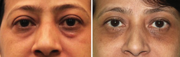
How Common Is Fat Repositioning in Contemporary Practice?
The review summarizes survey data from members of the American Society of Ophthalmic Plastic and Reconstructive Surgery. Nearly 96% of respondents reported using the transconjunctival approach, 82% reported using the transcutaneous approach, and 51% reported using both. About 80% performed fat repositioning. These numbers do not prove superiority, but they do demonstrate how strongly modern practice has moved toward flexible, anatomy-based and volume-preserving strategies.
The same survey reported different preferences for the plane of repositioning. Around 70% of respondents who repositioned fat preferred a supraperiosteal plane and 30% a subperiosteal plane. The review also cites a retrospective comparison in which supraperiosteal transposition was associated with more bruising, swelling and contour abnormalities than subperiosteal transposition. These apparently different findings illustrate an important point: surgeon preference, anatomy and technique details matter, and a survey of popularity is not the same as comparative evidence of best outcome.
What Is Canthopexy or Canthoplasty and Why Might It Be Added?
The lateral canthus contributes to the position and stability of the lower eyelid. If laxity is present before surgery, removing skin or altering the lower eyelid without adequate support can increase the risk of malposition. The review therefore discusses canthopexy and canthoplasty as integral adjuncts in selected lower eyelid procedures.
In the source article, minimal laxity of approximately 1–2 mm may be managed with a lateral canthal suture canthopexy, moderate laxity of approximately 3–6 mm may require a lateral retinacular canthopexy, and severe laxity greater than 6 mm may lead the surgeon to consider canthoplasty with cantholysis or a lateral tarsal strip. These thresholds come from the reviewed surgical literature and are not a self-assessment tool; lower-lid stability must be evaluated clinically.
The practical principle is that eyelid support should be treated as part of the rejuvenation plan rather than as an afterthought. A technically successful reduction of eye bags is not a good result if the lower eyelid is left retracted, rounded or unstable.
Integrated Lower Eyelid and Midface Approaches
Some patients have a lower eyelid problem that cannot be separated from the cheek. Flat malar projection, a deep tear trough, midface descent and lower-lid laxity may all contribute to the same tired appearance. The review describes integrated procedures combining lower blepharoplasty with SOOF manipulation, cheek lift, midface lift, septal techniques or lateral canthal support.
One described modification in patients with hypoplastic malar regions combines canthopexy and cheek lift with redraping of orbital fat and the SOOF to create a smoother lid-cheek transition. Another technique rotates a complex of capsulopalpebral fascia, orbital septum and orbital fat downward to camouflage the tear trough. These are examples of how lower eyelid surgery can be adapted to a three-dimensional structural problem rather than used as simple bag removal.
The source review is appropriately cautious: because the literature includes many different variants, there is not enough high-quality comparative evidence to identify one integrated method as the universally best option. The operative plan should therefore be based on anatomy, surgeon expertise and the balance of expected benefit and risk.

Can Lower Eyelid Bags Be Treated Without Surgery?
Not every patient with under-eye concerns requires blepharoplasty. The source review notes the increasing use of noninvasive or minimally invasive approaches such as laser resurfacing, dermal fillers and chemical peeling, as well as autologous fat grafting in selected circumstances. These treatments address different components of the problem.
For example, resurfacing or chemical peeling may improve skin quality but cannot reliably remove a substantial prolapsed orbital fat pad. Hyaluronic acid filler may soften selected hollows in patients whose main issue is volume loss or tear trough depression, but filler does not remove genuine skin redundancy and may be inappropriate in patients with certain patterns of fluid retention, severe bags or unfavorable anatomy. Fat grafting can restore volume but is also a separate procedure with its own risks and indications.
This distinction is particularly relevant for aesthetic clinics: “under-eye rejuvenation” is not one treatment. A patient may need skin-quality treatment, volume correction, surgery, or no intervention at all. The safest approach is to identify which anatomical component is creating the visible concern before selecting a modality.
Potential Risks and Complications
Lower eyelid blepharoplasty can produce durable aesthetic improvement, but it is surgery around the eye and should never be presented as risk-free. The source review identifies postoperative retro-orbital hemorrhage as the most alarming complication because it can threaten vision and requires immediate attention. Although rare, its seriousness reinforces the need for appropriate surgical training, hemostasis and postoperative monitoring.
Other complications described in the review include bruising, superficial hematoma, dry-eye symptoms, corneal trauma, infection, insufficient fat removal, injury to the inferior oblique muscle with diplopia, periocular pigmentation, asymmetry, lower eyelid retraction, scleral show, lagophthalmos, scarring and ectropion. Some problems are temporary, whereas others can require additional treatment or revision.
| Potential issue | Why it matters | Evidence-based context from the review |
|---|---|---|
| Retro-orbital hemorrhage | Rare but potentially sight-threatening | The review identifies this as the most alarming postoperative complication and emphasizes immediate attention. |
| Scleral show / retraction / ectropion | Can alter lower-lid position and ocular comfort | Reported more often with transcutaneous approaches in several cited series; risk varies with anatomy and technique. |
| Dry eye / lagophthalmos | May affect ocular surface symptoms | Preoperative dry-eye assessment and conservative tissue handling are important. |
| Diplopia from inferior oblique injury | Can impair binocular vision | The inferior oblique lies between medial and central fat pads and must be recognized and protected. |
| Hollowing / contour irregularity | May create an aged or skeletonized look | Complete fat removal can accentuate the tear trough; modern approaches often preserve and reposition fat. |
| Bruising, edema, hematoma, pigmentation | Common recovery concerns | The review lists these among frequently encountered postoperative issues; severity varies between patients and techniques. |
| Infection / corneal trauma | Uncommon but clinically important | Both are described among possible complications and require appropriate surgical and postoperative care. |
| Asymmetry / insufficient fat or skin correction | May affect aesthetic satisfaction | Revisions and residual contour issues are reported in some series, supporting conservative planning. |
What Do Published Series Say About Complication Rates?
The review collates complication data from several surgical series, but the numbers should not be compared as if they came from one controlled trial. Different studies used different patient populations, techniques, definitions and follow-up periods. Still, the data illustrate the types of events surgeons monitor.
- In one comparative dataset cited by the review, transconjunctival surgery was associated with a lower rate of scleral show than a transcutaneous approach.
- A series of 2,400 cases cited in the review reported ectropion, hollow eyes, dry eyes and retraction at low but non-zero rates, with retraction being the most frequent of those listed outcomes.
- Another series reported lower-lid malposition and revision procedures after an integrated lower blepharoplasty approach.
- Other reports included reoperation for skin ptosis, contour or pretarsal-roll asymmetry, sensory changes, hypertrophic scar, stitch sinus and chemosis.
- The existence of these complications supports conservative tissue handling, proper lower-lid support and careful patient selection.
For website communication, the most accurate message is not to quote a single universal complication percentage. Risk is affected by the chosen approach, the amount of tissue removed, the patient’s baseline lid tone, dry-eye status, canthal support, midface anatomy and the experience of the surgeon.
Recovery and Healing: What Can Be Said from This Review?
The source review does not provide one standardized day-by-day recovery schedule because the included techniques vary substantially. It notes that the conjunctival incision in a transconjunctival procedure usually heals within approximately one week, but swelling, bruising and contour settling are influenced by the extent of surgery and whether additional procedures are performed.
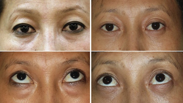
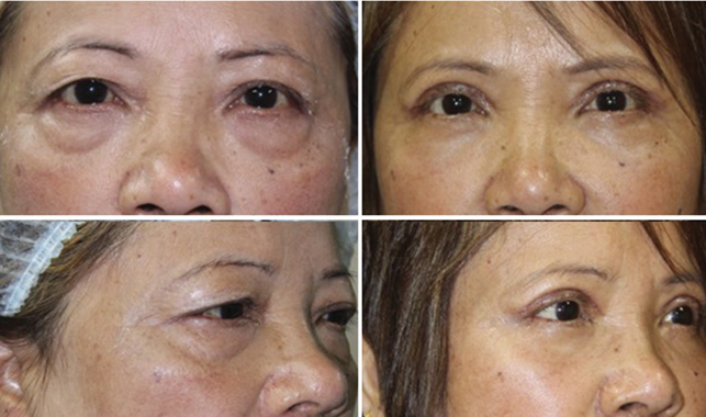
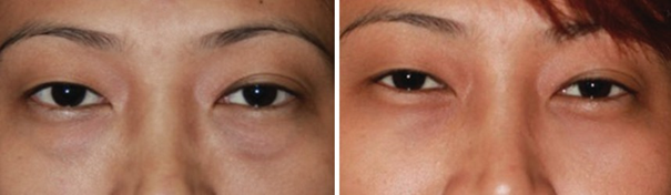
The clinical figures in the review show postoperative appearances at several different time points, including seven days, three months, six months and five years. These images are useful for demonstrating that early postoperative appearance is not the same as the final aesthetic result, but they should not be used to promise a fixed recovery timeline for every patient.
A patient-specific recovery discussion should consider the surgical route, fat excision versus repositioning, canthal support, midface work, skin excision, baseline ocular surface health and individual healing. Postoperative instructions and urgent warning signs should come directly from the operating surgeon.
What Do Long-Term and Integrated Results Look Like in the Source Review?
The authors report their experience with 540 patients and approximately 1,000 eyelids over 18 years, describing smooth effacement of the lower eyelid-cheek interface with transconjunctival lower eyelid blepharoplasty. They highlight the absence of external skin scarring and the ability of fat repositioning to improve the transition from the lower eyelid to the malar region.
A 6-month result after fat relocation with midface lift and lateral canthopexy, a 3-month result after lower lid blepharoplasty and a 5-year postoperative comparison. These images are individual examples rather than guarantees of outcome. Their value is in illustrating the central aesthetic objective of contemporary lower blepharoplasty: not simply removing a “bag,” but creating a balanced and smooth lower-lid contour.
What the Evidence Does Not Prove
The review is explicit about the limitations of the literature. Studies use multiple surgical variations, not all papers describe the exact fat-transposition technique, and there is no general agreement on which procedure is best for a given age or ethnicity. The article also notes the lack of left-right comparative lower-blepharoplasty studies in which one side receives one technique and the other side receives another.
This matters because aesthetic surgery can be strongly influenced by surgeon experience and patient selection. A technique may appear superior in one series because it was used in a different type of patient. For this reason, high-quality consultation and anatomy-based planning remain essential even when a procedure is widely used.
The strongest conclusion from the review is therefore not “transconjunctival is always best” or “fat repositioning is always necessary.” Instead, the evidence supports conservative tissue management, preservation of orbital fat when appropriate, careful recognition of lower-lid support, and an individualized choice between transconjunctival, transcutaneous and integrated approaches.
A Practical Decision Framework for Patients
A useful way to think about lower eyelid treatment is to separate the visible complaint into anatomical components. The following framework is educational and is not a substitute for an examination :
- Is the main problem a true bulge from orbital fat, or is the lower eyelid mainly hollow?
- Is there genuine excess skin that needs surgical removal, or is the skin simply thin, crepey or photodamaged?
- Is the tear trough prominent because of volume loss, fat prolapse above it, skeletal support, or a combination?
- Is the lower eyelid tight and stable, or is there horizontal laxity and canthal weakness?
- Is the cheek projected enough to support a smooth lower lid-cheek transition?
- Are festoons or malar mounds part of the appearance?
- Are there dry-eye symptoms, previous eyelid surgery or other ophthalmic factors that alter the risk profile?
- Would a non-surgical treatment address the actual problem, or would it camouflage rather than correct it?
Once these questions are answered, the surgical route becomes more logical. Isolated fat prolapse with good skin can point toward a transconjunctival strategy. Meaningful skin redundancy can justify a transcutaneous component. A pronounced tear trough can favor fat preservation or repositioning. Lateral laxity can require canthal support. Midface deficiency may require an integrated approach. The best plan is therefore a diagnosis-driven combination, not a one-size-fits-all package.
