
Why the Periocular Area Ages Early
The eye region is one of the earliest areas of the face to show age-related change. The source describes brow descent, loss of connective-tissue support, upper-eyelid laxity, lateral hooding and a tired appearance as interrelated features rather than isolated problems.
This is an important clinical distinction. A patient may describe “heavy eyelids,” but the visible redundancy can partly reflect descent of the brow and adjacent tissues. Likewise, lower-eyelid puffiness and fine lines may sit within a broader continuum that includes the lateral canthus, temple and infraorbital support.
Because these structures function together, the study questioned whether treating only the eyelid is too narrow an approach for selected patients with early periocular laxity.
The Expanded Periocular Concept
The defining feature of this study was not simply the use of radiofrequency; it was the decision to treat the periocular complex as an integrated anatomical unit. The expanded field included the suprabrow forehead, upper eyelid, lateral canthus, temple and infraorbital region.
This framework attempts to address adjacent structural contributors to the eye-area appearance. For example, the source notes that brow descent can worsen upper-eyelid redundancy and lateral hooding. A broader treatment plan may therefore create a more harmonious change than a strictly eyelid-only pattern.
The concept should still be individualized. Expanded-field treatment does not mean every square centimeter around the eye receives the same energy, depth or treatment tip.
How XERF Dual-Frequency Monopolar RF Works
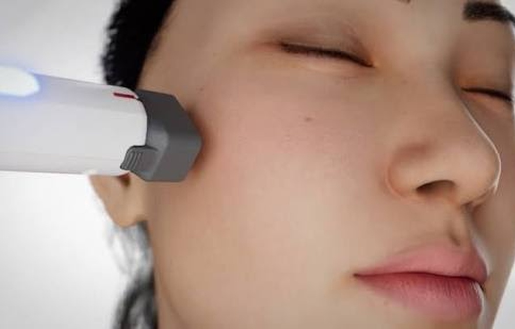
The study used XERF (Cynosure Lutronic), a monopolar RF platform operating at 6.78 MHz and 2 MHz. The article describes monopolar RF as producing controlled volumetric heating through tissue impedance.
Thermal exposure can create immediate collagen contraction followed by longer-term neocollagenesis and elastin remodeling. The expected clinical effect is therefore progressive tissue contraction and remodeling rather than an ablative removal of skin.
The source also notes that RF is chromophore-independent. In practical terms, the energy is not dependent on melanin absorption in the way many light-based technologies are. This characteristic is one reason RF has been investigated in a broader range of skin types, although careful periocular technique remains essential.
Why Two Small Effectors Were Used
The periocular region is anatomically heterogeneous. The upper eyelid is thin and sensitive, while the suprabrow forehead and temple have thicker soft-tissue coverage. The study therefore used two different small RF effectors rather than one uniform applicator.
Effector05 measured 5 × 10 mm and was primarily applied to the upper eyelid and other narrow, delicate zones requiring precise localized energy delivery.
Effector10 measured 10 × 10 mm and was used along the orbital rim and in broader structural zones such as the suprabrow forehead, temple and infraorbital region. The authors relate this choice to the principle that electrode geometry can influence current density and tissue penetration.
| Study effector | Size | Primary source-described use | Clinical logic |
|---|---|---|---|
| Effector05 | 5 × 10 mm | Upper eyelid and adjacent precision areas | Focal delivery in thin, curved and sensitive tissue |
| Effector10 | 10 × 10 mm | Suprabrow forehead, temple, orbital rim, infraorbital region | Broader volumetric heating in thicker structural zones |
Study Design and Patient Population
This was a retrospective exploratory pilot study rather than a randomized controlled trial. Fourteen patients with clinically evident mild-to-moderate periocular laxity were included and all completed the approximately three-month follow-up.
The cohort consisted of 12 women and 2 men. Mean age was 53.1 ± 8.0 years, with a range of 40 to 67 years. Eight patients had Fitzpatrick skin type III and six had type IV.
The inclusion profile is important because the study was designed around early-to-moderate laxity. It should not be extrapolated automatically to severe brow ptosis, major dermatochalasis, large fat prolapse or patients who already meet surgical indications.
| Baseline characteristic | Study value |
|---|---|
| Number of patients | 14 |
| Female / male | 12 (85.7%) / 2 (14.3%) |
| Mean age | 53.1 ± 8.0 years |
| Age range | 40–67 years |
| Fitzpatrick III | 8 (57.1%) |
| Fitzpatrick IV | 6 (42.9%) |
| Laxity severity | Mild-to-moderate periocular tissue laxity |
Who Was Excluded from the Study ?
The source excluded pregnancy or breastfeeding, active dermatologic or ocular disease in the treatment area, impaired wound healing, a history of keloid formation, and surgical or energy-based periocular procedures during the preceding six months.
These exclusions describe the study population; they should not be treated as a complete universal contraindication list. Current device instructions, medical history, eye history and local clinical protocols remain necessary for individual treatment decisions.
The Source-Specific Treatment Protocol
Each participant underwent one XERF session. RF energy was delivered in multiple overlapping passes across the expanded periocular field.
The study reports a total of 400–900 shots per treatment, distributed according to anatomical coverage, tissue thickness and degree of laxity. Mean total delivered energy was 6.44 ± 3.16 kJ per session.
Treatment parameters were adjusted according to regional sensitivity and patient tolerability. This individualized approach is particularly important around the eye, where tissue thickness can change substantially over a very short distance.
Ocular Protection Was a Core Safety Step
The study used non-metal protective intraocular eye shields during treatment. Direct pressure over the globe was avoided, and continuous patient feedback was obtained to monitor discomfort or visual disturbance.
This safety detail is important because “non-invasive” does not mean that the eye area can be treated casually. The authors specifically emphasize that periocular anatomy is sensitive and that appropriate ocular protection is necessary during energy-based procedures.
No ocular adverse events were reported in the 14-patient cohort, but a small cohort cannot establish that risk is zero.
How the Study Measured Results
Standardized photographs were obtained at baseline and approximately three months after treatment under identical lighting, camera settings and positioning.
Two independent blinded evaluators who were not involved in the procedures assessed improvement using the Global Aesthetic Improvement Scale (GAIS). On this 5-point scale, 1 meant very much improved, 2 much improved, 3 improved, 4 no change and 5 worse.
Patient satisfaction was assessed separately with a 5-point Likert scale. Pain and treatment-related adverse events were also recorded.
What Visibly Improved ?
The source reports bilateral improvement in periocular appearance in all treated subjects. Clinically visible changes included less upper-eyelid heaviness, improvement in eyelid hooding, decreased lower-eyelid puffiness, sharper definition of the upper-eyelid crease and reduction of fine periocular wrinkles.
These outcomes are consistent with the broader treatment concept: the study was not attempting to create one isolated eyelid effect, but to improve the visual relationship between the brow, eyelid, lateral canthus, temple and infraorbital region.
Importantly, the study did not provide an objective millimeter measurement of brow elevation, eyelid skin reduction or lower-eyelid volume. The visible changes should therefore be described as photographic/clinical improvement rather than a quantified surgical-equivalent lift.

Patient Satisfaction
Patient-reported satisfaction was high. The mean satisfaction score was 4.36 ± 0.63 on a 5-point scale, with a median of 4 and an interquartile range of 4–5.
Thirteen of 14 participants (92.9%) reported high satisfaction, defined as a score of 4 or 5. Six of 14 (42.9%) rated themselves “very satisfied.” No patient reported dissatisfaction.
These are encouraging figures, but they belong to a small selected cohort and should not be converted into a universal expected satisfaction rate for every XERF eye-area patient.
Pain and Comfort
Mean pain score was 2.33 ± 0.75 on the study’s 5-point scale. In the discussion, the authors describe the procedure as well tolerated and note low pain despite the absence of anesthesia.
Low average pain in a 14-patient study does not mean the procedure is painless for everyone. Sensitivity varies substantially around the eyes, and patient comfort can depend on anatomy, settings, number of passes and individual pain threshold.
Downtime and Short-Term Recovery
Some patients developed transient mild swelling of the upper eyelid during the first post-treatment week. The study reports no clinically significant persistent erythema, discomfort, functional impairment or ocular complications during follow-up.
This makes “minimal downtime” a reasonable description of the cohort. “Zero downtime,” however, would be less precise because mild temporary swelling was specifically observed.
Safety Results
No burns, post-inflammatory hyperpigmentation or ocular complications were reported. The study also did not report clinically significant persistent discomfort or functional impairment.
The cohort predominantly included Fitzpatrick skin types III and IV. The absence of pigmentary complications is encouraging in this context, but only 14 patients were studied and uncommon complications could easily be missed.
A good evidence-based page should therefore say that the procedure was well tolerated in this pilot study rather than describing it as risk-free.
| Safety / tolerability measure | Source finding |
|---|---|
| Mean pain score | 2.33 ± 0.75 / 5 |
| Transient swelling | Mild upper-eyelid swelling in some patients during first week |
| Clinically significant erythema | None reported |
| Burns | None reported |
| Post-inflammatory hyperpigmentation | None reported |
| Ocular complications | None reported |
| Functional impairment | None reported |
Why Skin Type Matters
Laser resurfacing can be effective for superficial periocular rhytides, but the source notes that pigmentary risk and downtime can be important considerations, especially in higher Fitzpatrick skin types.
Monopolar RF is described as chromophore-independent, which means melanin is not the primary target of the energy. That feature may reduce pigment-related constraints compared with some light-based procedures, although it does not eliminate all risks or remove the need for conservative periocular treatment.
This study included only Fitzpatrick III and IV patients; it does not establish identical safety or efficacy across every skin type.
Who Appears to Fit the Study Profile Best ?
The source is most applicable to adults with mild-to-moderate periocular laxity who want gradual, non-surgical improvement and minimal recovery time.
The visible study targets included upper-eyelid hooding/heaviness, early brow-related laxity, lateral periocular laxity, infraorbital puffiness and fine wrinkles.
Patients with advanced excess upper-eyelid skin, severe brow ptosis, large lower-eyelid fat prolapse, major functional visual-field obstruction or other ocular pathology were not the focus of this study and should not be assumed to have equivalent outcomes.
XERF vs Blepharoplasty : Different Levels of Correction
The source acknowledges blepharoplasty as an effective corrective procedure. Surgery can physically remove or reposition tissue and remains a different category of treatment from non-invasive RF.
XERF in this study was evaluated as an early-stage, maintenance-oriented or prejuvenation option for mild-to-moderate laxity. It should not be marketed as a substitute for surgery in patients with major structural excess or functional eyelid problems.
A consultation should therefore answer the diagnosis first: is the problem early laxity that may respond to remodeling, or is there enough excess tissue or ptosis to require surgical evaluation?
XERF vs Laser Resurfacing
Laser resurfacing primarily targets skin texture and superficial rhytides through a different tissue interaction. The source mentions that periocular laser use can be constrained by downtime and pigmentary concerns in some skin types.
The 2026 XERF study did not directly compare dual-frequency RF with fractional laser or ablative laser. No superiority claim can be made from this paper. In practice, the two categories may address different dominant problems.
XERF vs HIFU and Other Tightening Devices
The uploaded paper does not compare XERF directly with HIFU, microfocused ultrasound, other monopolar RF systems, RF microneedling or surgical lifting.
Therefore, statements such as “better than HIFU” or “better than Thermage” are not supported by this study. The value of this paper is narrower and more defensible: it documents a specific expanded-field XERF approach around the eyes with favorable short-term photographic, satisfaction and safety outcomes.
When Should Results Be Judged ?
Formal outcome assessment occurred at approximately three months. That timing matches the biologic rationale described by the authors: thermal collagen contraction can occur early, but neocollagenesis and elastin remodeling develop progressively.
The study cannot establish the full long-term durability of the result because it did not report six-, twelve- or eighteen-month follow-up. Any durability claim beyond the three-month study window would require separate evidence.
XERF Eye Tightening in Abu Dhabi with Dr. Erfan Rahmani
For patients researching XERF for the eye area in Abu Dhabi, the central question is not simply whether XERF can be used around the eyes. It is whether the visible concern is the type of early laxity and tissue change that a non-invasive RF strategy can realistically improve.
Dr. Erfan Rahmani can assess brow position, upper-eyelid hooding, lateral periocular support, infraorbital tissue, skin quality and whether a device-based plan is appropriate or whether another treatment category should be considered.
More Information : www.drerfanrahmani.com
