

Hair Loss Treatment Starts With Understanding The Cause
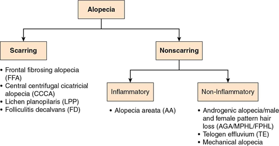
Many people who seek hair restoration have androgenetic alopecia (male- or female-pattern hair loss), but it is important not to assume that every case of thinning is the same. Other conditions can cause diffuse shedding, patchy hair loss, inflammation or permanent scarring. The first goal of a consultation is therefore to identify the type of alopecia and determine whether cosmetic hair-restoration treatment is appropriate.
This matters because treatments that may be suitable for ordinary pattern hair loss may not address an active inflammatory, autoimmune, nutritional, hormonal or scarring process. In some cases, investigating the underlying cause is the most important first step.
Understanding The Normal Hair Cycle
Hair does not grow continuously. Each follicle moves independently through a growth phase (anagen), a short transition phase (catagen) and a resting/shedding phase (telogen). Most scalp follicles are normally in the growth phase, while a smaller proportion are resting or shedding at any given time.
Because the hair cycle is measured in months and years rather than days, visible improvement from many hair-loss treatments also takes time. This is why consistent follow-up and standardized photographs are more useful than judging progress from one week to the next.

Common Causes Of Hair Loss That Can Look Similar
1. Androgenetic Alopecia : Male & Female Pattern Hair Loss
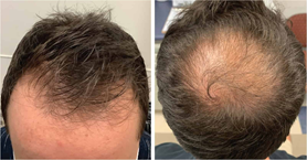
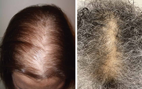
Androgenetic alopecia (AGA) is the most common form of hair loss in adults. Over time, affected follicles become progressively smaller, a process called miniaturization. The hair shaft becomes thinner and the growth phase becomes shorter.
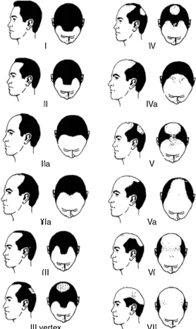
In men, thinning commonly involves the temples, frontal scalp, mid-scalp and crown. In women, the pattern is often more diffuse over the crown and central scalp, although several distribution patterns can occur. The back of the scalp is often relatively preserved in classic AGA.

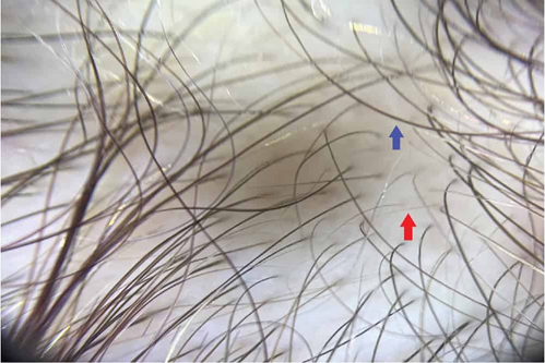
2. Telogen effluvium: sudden or increased shedding
Telogen effluvium (TE) usually presents as diffuse shedding rather than a sharply defined bald patch. Patients may notice much more hair in the shower, on a brush or on the pillow. It can follow a physical or emotional stressor several months earlier.
- Severe illness, hospitalization or major surgery.
- Major emotional stress.
- Childbirth or hormonal medication changes.
- Significant weight loss or crash dieting
- Thyroid disorders, anemia or iron deficiency
- Certain medications
Telogen effluvium and pattern hair loss can occur at the same time. A sudden episode of shedding may make previously subtle pattern thinning much more noticeable.
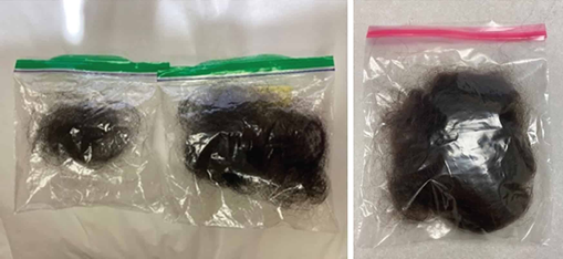
3. Alopecia Areata : Patchy Autoimmune Hair Loss
Alopecia areata is an autoimmune, non-scarring form of hair loss. The classic presentation is the sudden appearance of one or more well-defined smooth patches, but the condition can also be diffuse or extensive. Eyebrows, eyelashes or beard hair may also be involved.
Dermoscopy can reveal characteristic short, tapered “exclamation-mark” hairs in active disease. These findings help distinguish alopecia areata from ordinary pattern hair loss.
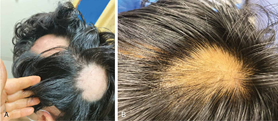
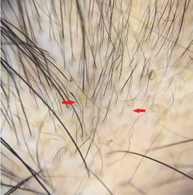
4. Mechanical or Traction-Related Hair Loss
Repeated pulling forces from tight hairstyles can damage follicles, particularly around the front and temples. Early traction alopecia may be non-scarring and potentially reversible, but long-standing traction can become scarring and permanent. Irregular or geometric patches can also occur in hair-pulling disorders.
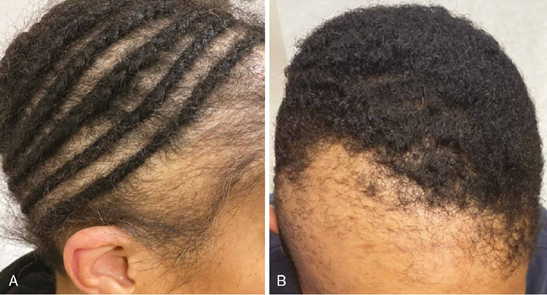
When Hair Loss May Be Scarring
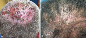
Scarring alopecia is less common than ordinary pattern hair loss, but it is especially important to recognize because the follicle can be permanently destroyed and replaced by scar tissue. Early diagnosis may help prevent additional irreversible loss.
Features that Deserve Closer Medical AssessmentItching, burning, scalp pain, redness, scaling, follicular bumps or pustules, smooth shiny areas with loss of follicular openings, progressive eyebrow loss, or an unusual distribution of hair loss can raise suspicion for an inflammatory or scarring condition.
Examples of Scarring Alopecia Patterns
Lichen planopilaris may present with perifollicular scale, redness, keratotic plugs, burning, tenderness or itching. Frontal fibrosing alopecia often causes progressive frontotemporal recession and may be associated with loss of eyebrows or sideburn hair.
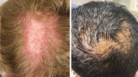
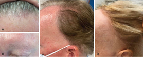

Central centrifugal cicatricial alopecia typically begins around the crown or vertex and expands outward. Folliculitis decalvans can cause recurrent painful pustules, crusting and scarring with tufted hairs.
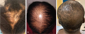

What Happens During a Hair-Loss Consultation?
A useful hair-loss consultation is more than a quick look at the scalp. The aim is to connect the pattern of hair loss with the patient’s timeline, symptoms, medical history, medications, nutrition, hormonal history and previous treatments.
| 1. Clarify the main problem | Is the concern increased scalp visibility, thinning, active shedding, or a focal bald patch? Was the onset sudden or gradual? |
| 2. Review recent triggers | Important events can include severe illness, surgery, psychological stress, childbirth, medication or hormonal changes, and major weight change. |
| 3. Assess risk factors | Nutrition, endocrine disease, iron deficiency, autoimmune history, previous cancer therapy, medications and relevant reproductive or hormonal symptoms may influence the assessment. |
| 4. Examine the scalp and hair pattern | The distribution, severity, presence of inflammation, scarring, broken hairs, pustules or scaling are assessed. Eyebrows, eyelashes, beard and other hair-bearing areas may also provide diagnostic clues. |
| 5. Use dermoscopy when appropriate | Magnified examination can identify follicular miniaturization, hair-diameter variation, inflammatory changes, missing follicular openings and other features that are difficult to see with the naked eye. |
| 6. Decide whether additional tests are needed | Laboratory testing or scalp biopsy is not required for every patient; it is guided by the history and examination. |
Why Dermoscopy Can Be Useful
Dermoscopy provides a magnified view of the scalp and follicular openings. In androgenetic alopecia, one of the key findings is variation in hair-shaft diameter due to miniaturization. Dermoscopy can also reveal signs of inflammation, scarring, alopecia areata or mechanical damage, helping the clinician decide whether the picture fits ordinary pattern hair loss or requires a different workup.
Do all patients need blood tests ?
No. Testing should be guided by the history and examination. When an underlying medical contributor is suspected, commonly considered investigations include a complete blood count, iron studies such as ferritin, and thyroid testing. Other tests may be selected when there are specific clues, such as hormonal symptoms, dietary restriction, autoimmune symptoms or relevant infection risk.
Important NuanceLow iron and vitamin D levels are commonly discussed in hair-loss care, but the source material notes that the strength of the relationship is not equally established for every patient. Testing and supplementation should therefore be individualized rather than automatic.
When Might a Scalp Biopsy be Considered ?
A scalp biopsy can be valuable when the pattern is atypical, when symptoms or examination raise concern for inflammation or scarring, or when the diagnosis remains uncertain. This is particularly important in suspected scarring alopecia because early diagnosis can affect the chance of preventing further permanent follicular loss.
How Treatment Progress Should Be Monitored
Hair-restoration treatment often requires long-term follow-up. Standardized scalp photography and, when available, videodermoscopy can make changes easier to measure objectively. Consistent lighting, angle, magnification and hair positioning improve the usefulness of before-and-after comparisons.
- Frontal view
- Vertex/crown view
- Occipital (back of scalp) view
- Right and left temporal views
- Magnified dermoscopic images when clinically useful
When Should You Consider a Medical Hair-Loss Assessment ?
- Hair loss started suddenly or shedding has increased significantly.
- You have patchy bald areas rather than gradual patterned thinning.
- The scalp is itchy, painful, burning, red, scaly or develops pustules/crusting.
- There is eyebrow, eyelash or beard loss in addition to scalp hair loss.
- Hair loss followed major illness, surgery, childbirth, a new medication or major weight change.
- You are considering PRP, regenerative treatments, medication or hair transplantation but the diagnosis has not been clearly established.
- Previous hair-loss treatment has not produced the expected result.
