
Dermal Filler Complications : Complete 2026 Guide to Signs, Risks and When to Seek Help
Quick answer : What are the main complications of dermal fillers?
Dermal fillers can produce predictable short-lived effects such as swelling, tenderness and bruising. They can also cause poorly placed product, visible lumps, delayed inflammatory nodules, infection, allergic reactions and injury to nerves or nearby structures. The most serious uncommon events are vascular occlusion, skin ischemia or necrosis, vision loss and stroke. A complication may emerge immediately, after several days, or even months after treatment. Early recognition matters because some serious complications are time-critical.[1–6]
The central principle is simple: not every lump is an infection, not every bruise is an occlusion, and normal-looking surface skin does not reliably exclude deeper injury. Safe decisions depend on symptoms, progression, examination, exact product and treatment history, anatomical location and, where appropriate, diagnostic ultrasound and specialist assessment.
The difference between an expected side effect and a complication
A mild, improving bruise or short-lived swelling after injection may be within the anticipated post-treatment course. A complication is an unwanted event that requires closer evaluation or alters the expected course. These categories overlap: a bruise can be cosmetically distressing, a hematoma can exert pressure on adjacent structures, and painful swelling can be an early sign of something considerably more serious.
| Presentation | Often compatible with routine recovery | Requires prompt medical review or emergency response |
|---|---|---|
| Swelling | Mild and gradually settling | Rapidly worsening, unusually painful, associated with fever or airway symptoms |
| Bruising | Localized discoloration evolving over days | Expanding hematoma, tense swelling, associated neurologic or ischemic signs |
| Lump | Small, painless, stable early irregularity | Red, hot, tender, enlarging, recurrent, late-onset or unusually firm lesion |
| Skin color | Minor redness after puncture | Blanching, livedoid/mottled appearance, grey/dusky change, rapidly progressive discoloration |
| Sensation | Temporary local tenderness | Severe out-of-proportion pain, persistent numbness or weakness |
| Vision / neurologic symptoms | None expected | Any visual symptom or new stroke-like symptom is an emergency |
This table is a triage aid, not a method of diagnosing a complication at home. If a symptom feels unusual, the safest choice is to contact the treating medical professional promptly. If vision, circulation or neurologic function may be affected, seek immediate emergency care.
Why dermal filler safety is fundamentally an anatomy problem
The face contains complex arteries, veins, sensory and motor nerves, muscles, fat compartments, salivary glands, orbital structures and tight connective-tissue spaces. Vessels communicate across regions, and individual anatomy varies. Injected material can distort tissue, be compressed by swelling or, in rare cases, enter an artery and travel to downstream tissues. The consequences depend on both anatomy and the type of filler.
Arteries, ischemia and embolization
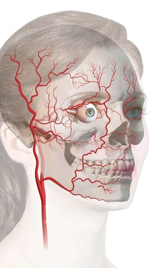
An accidental intravascular injection can block local circulation. Some vessels link facial injection territories to the ophthalmic circulation, which explains how blindness is possible after injection at sites that are not physically inside the eye. Neurological injury may follow embolization into the cerebral circulation. The FDA specifically identifies skin necrosis, blindness and stroke among rare but serious consequences of intravascular filler administration.[1]
Why the nose and upper face deserve special caution
The nose has relatively constrained spaces, a complex arterial supply and delicate soft tissues. The glabella, forehead and periorbital regions have relevant vascular connections. A small cosmetic change does not imply a small medical risk. The safety profile of a specific treatment also depends on the exact product, technique, practitioner training and patient factors. U.S. FDA approvals do not cover every facial injection site, and other jurisdictions have their own requirements.[1]
Anatomical structures beyond vessels
Needles and cannulas may affect nerves and salivary structures, particularly around the chin, jawline and parotid region. A rare injury can resemble a more common post-treatment swelling or inflammatory reaction. Knowledge of layered anatomy, not reliance on a single advertised "safe plane," is therefore essential.
1. Non-inflammatory filler lumps : why a painless bump develops

Non-inflammatory lesions, including lip irregularities, usually present as palpable or visible bumps without marked redness, heat or tenderness. Possible explanations include product placement, uneven distribution, a discrete collection of filler, superficial placement, migration, localized swelling or a benign pre-existing structure noticed after treatment.
Small early irregularities may improve as tissue edema resolves. Persistent cosmetic asymmetry, increasing size or uncertainty about the nature of a lump warrants clinical review. An injector must also distinguish hyaluronic-acid products from calcium hydroxyapatite, poly-L-lactic acid, permanent materials and other fillers, because management is not interchangeable.
Should you massage a lump after lip filler?
Not automatically. Gentle massage is sometimes discussed for uncomplicated non-inflammatory lip lumps, but that should never be taken as a blanket instruction. Pressure on an ischemic or infected region can obscure assessment and delay appropriate care, and superficial products, firm nodules and delayed lesions may require entirely different management. Only massage a treated area when the responsible clinician has examined the problem and specifically advised it.
When can ultrasound change the diagnosis?
High-frequency ultrasound can help determine whether a palpable abnormality represents filler, edema, inflammatory tissue, abscess, foreign material, vascular change or another structure. A 2025 international Delphi consensus recommends specific training, Doppler capability and ultrasound use for important aesthetic-complication scenarios. It is not a handheld gadget that makes every injection safe.[5]
2. Lumps in the lips : immediate, early and delayed reactions
Lip concerns deserve a dedicated diagnostic approach, because normal lip anatomy, high mobility, repeated injection history and patient expectations complicate interpretation. Relevant history includes the product name and amount, injection date, previous fillers, location of the lump, onset, medication, dental treatment, recent infection, allergy history and whether there were similar reactions previously.
Early lip lumps
Within hours to days, edema, bruising, contour irregularity or an early inflammatory reaction may be responsible. Progressive pain, blanching or unusual color change immediately after injection is not a routine "lump" and must trigger urgent vascular assessment. Diffuse swelling may evolve for a short period, but deterioration rather than improvement should be reviewed.
Delayed lip nodules
Weeks, months or occasionally years later, a nodule may represent persistent product, a delayed inflammatory response, a granulomatous reaction, or infection including possible biofilm involvement. Firm recurrent swellings and episodes of tenderness are typical descriptions. Modern consensus reviews recognize several interacting hypotheses (product properties, microbial factors and patient immune response) rather than a single proven cause for every delayed nodule.[6]
What a structured consultation should establish
A clinician should record the material involved if known, its injection location and date, whether the nodule is solitary or multiple, whether it is fluctuant or solid, the degree of pain and erythema, any fever or mucosal findings and previous attempted treatments. Ultrasound can be helpful when product identity or depth is uncertain. If infection is suspected, indiscriminate treatment without a diagnosis can delay proper care.
3. Inflammatory lesions and delayed-onset nodules
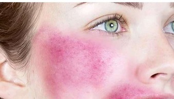
An inflammatory nodule is more than a cosmetic bump. It can arise from local tissue response, hypersensitivity, infection, post-procedural inflammation or a combination. Typical features include tenderness, warmth, redness, intermittent swelling, firmness or recurrence. Timing is useful but not definitive: an infection can appear early, and an immune-mediated reaction can appear well after the original procedure.
Differential diagnosis
- Infection: symptoms may include increasing warmth, tenderness, erythema, discharge, fever or a fluctuant collection. An abscess may require imaging and urgent management.
- Inflammatory foreign-body response: delayed firm or recurrent nodules may reflect inflammatory activity around material. "Granuloma" is a specific histologic diagnosis and should not be used as a synonym for every lump.
- Hypersensitivity: localized or diffuse swelling can reflect a reaction to product components or other exposures. Rare severe allergic symptoms require emergency treatment.
- Product accumulation or migration: non-inflammatory deposits can sometimes be mistaken for inflammation. Ultrasound is useful when location or product type is unclear.
The 2024 CARE consensus emphasizes medical history, aseptic practice, assessment of previous filler, recent infectious or dental events, and individualized investigation and care. A clinician should not automatically prescribe steroids to a possibly infected lesion, dissolve all nodules without identifying the material, or assume that every fluctuation is an allergy.[6]
Why empathy and documentation are medical necessities
Even a small visible lump can affect a person's willingness to smile, eat socially, attend work or appear in photographs. Explain uncertainty clearly, document the lesion objectively, arrange timely follow-up and avoid dismissing concerns. Reassurance is appropriate only after important red flags have been considered.
4. Bruising and hematoma after dermal fillers

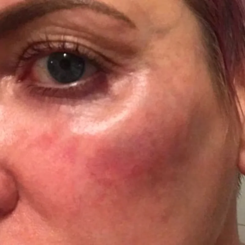
Ecchymosis describes discoloration caused by blood leaking into tissues, while a hematoma is a localized collection of blood. Petechiae and purpura are related descriptive terms. Bruising may be common after injection, but it is still meaningful: it can cause pain, unwanted visibility, anxiety and lost confidence in treatment.
Patients may feel embarrassed about conspicuous bruises, particularly around the eyes. Contemporary care includes expectation setting before treatment, respectful aftercare and documenting changes. The severity and expansion of a hematoma matter more than reassurance based on its presumed cosmetic nature.
Bruise or vascular occlusion?
Both can show discoloration. A bruise usually follows bleeding into tissue and evolves through recognizable color changes, whereas ischemia may present with severe pain, blanching, abnormal capillary refill, cool skin or livedoid patterns. These signs are not perfectly sensitive: normal coloration, preserved capillary refill or absent severe pain does not reliably exclude a deeper vascular injury. If concern remains, urgent assessment is required.
Sensible patient advice
Follow individualized written aftercare instructions and do not change prescribed blood-thinning medication independently. New expanding swelling, marked asymmetry with pain, compromised vision, neurologic symptoms or breathing symptoms require prompt escalation. Do not attempt to drain a hematoma at home.
5. Bacterial infection, cellulitis, abscess and biofilm-associated concerns

Bacterial infections after filler injections are generally uncommon but potentially serious. They range from a small local infection to cellulitis, abscess or systemic illness. Skin preparation, sterile equipment, careful technique and wound care all matter, including the risk of microorganisms entering around the lips or oral commissures.
What infection can look like
Look for increasing warmth, tenderness, spreading erythema, pus, fever, progressive swelling or feeling unwell. Some late infections can be subtle and intermittent. A localized lesion should not automatically be labeled a biofilm infection: the term refers to microbial communities that are difficult to prove clinically in an individual case. A differential diagnosis remains essential.
What assessment may involve
A medical review may include physical examination, ultrasound to identify a collection, cultures when there is drainage or an appropriate sample, review of immune status, and specialist advice for persistent or atypical infection. A suspected abscess is not equivalent to an uncomplicated non-inflammatory nodule. Treatment decisions depend on the actual presentation, product and patient.
Aseptic practice and follow-up
Prevention extends beyond pre-injection antisepsis. Proper skin preparation, sterile equipment, appropriate clinical facilities, avoidance of injecting through active infection, clear instructions for hygiene and a reachable follow-up service all matter. Patients should be told whom to contact if symptoms develop after office hours.
6. Nerve injury : neuropraxia, axonotmesis and neurotmesis

Aesthetic injections can rarely injure sensory or motor nerves through mechanical trauma, compressive swelling or indirect tissue injury. Three nerve-injury categories are commonly distinguished:
- Neuropraxia: a conduction disturbance, often transient.
- Axonotmesis: axon injury with supporting structures variably preserved.
- Neurotmesis: major disruption with poorer recovery potential.
These are clinical concepts, not labels that can be assigned reliably from a selfie or short video. Symptoms may include focal numbness, tingling, neuropathic pain, altered smile, lip asymmetry or weakness. Facial weakness may also result from anesthesia, swelling, ischemia or unrelated neurologic illness. A new facial droop with speech problems, limb weakness or visual change must be treated as a possible stroke, not as a routine post-filler nerve complaint.
Viral reactivation is a separate process. A herpes simplex outbreak around the lip should not be conflated with varicella-zoster-related neuritis or a directly punctured nerve. These are distinct diagnoses and need appropriate medical evaluation.
Why recovery cannot be promised at consultation
The timing and likelihood of recovery depend on the site and mechanism of injury, severity, time to recognition and any associated tissue ischemia. Avoid guaranteeing a particular recovery period. Document neurological findings and arrange a specialist referral when indicated.
7. Salivary gland injury during chin and jawline filler treatment
Jawline contouring brings instruments near the parotid and submandibular salivary glands, ducts, the facial nerve and layered fascia. Cannulas can still cause harm: a blunt tip does not grant immunity from tissue penetration or compression.
A gland injury may present with swelling, discomfort, altered salivation, localized tenderness or a persistent collection. These findings may resemble infection or ordinary post-injection edema. The clinical history, location and targeted imaging are important, and some cases require assessment by a maxillofacial or ear, nose and throat specialist.
The limitation of simplified "safe depth" diagrams
A diagram can help explain the relative relationships of dermis, fat, fascia, gland and muscle, but it cannot guarantee depth or safety in an individual face. Facial fat thickness, previous surgery, changes from aging and gland size vary. Real-time anatomical interpretation and appropriate competence are required.
8. Blindness after dermal filler: rare, devastating and time-critical
Can dermal fillers cause blindness? Yes. Although rare, filler injected into the facial vascular system may interrupt blood flow to ocular tissues. Filler-related vision loss has been reported after injections in several facial regions. A patient may report sudden blurred vision, a dark area in the visual field, complete loss of sight, eye discomfort or unusual headache. Associated neurologic symptoms are especially concerning.[1,4]
Early publications and historical compilations of published blindness cases establish why practitioners must understand vascular connections. Older case counts must not be mistaken for current worldwide incidence or a patient's exact risk. Case reports are vulnerable to reporting bias, and safe practice cannot be reduced to a single risk percentage.
How can injection outside the eye affect vision?
The facial arterial network communicates with the ophthalmic arterial system. Under certain injection conditions, filler may enter a vessel, move against normal flow under injection pressure and subsequently travel toward the retinal or ophthalmic circulation. Ocular ischemia can occur without the needle ever entering the orbit, and associated cerebral events have also been described. The mechanism is primarily arterial embolization. Older simplified descriptions of blockage in "an artery or vein" should not be presented as equivalent established mechanisms.
What should happen if visual symptoms begin?
The newer UK multidisciplinary consensus emphasizes transfer without delay, awareness of local emergency facilities, explicit consent covering the rare risks of vision loss and stroke, and preparedness before treatment begins. It also discusses specialized measures that qualified clinicians may consider, but their evidence is limited and none should delay hospital transfer.[4]
Does hyaluronidase always restore sight?
No. Hyaluronidase is used by qualified clinicians for hyaluronic-acid-related problems and is central to established skin-ischemia protocols, but evidence for restoring vision after an embolic ocular event is much less certain. Intrabulbar or retrobulbar interventions carry additional risks and are not a home or routine-clinic treatment. Emergency care must be guided by specialist pathways, product type and the situation.
9. Impending skin necrosis and filler-induced vascular occlusion
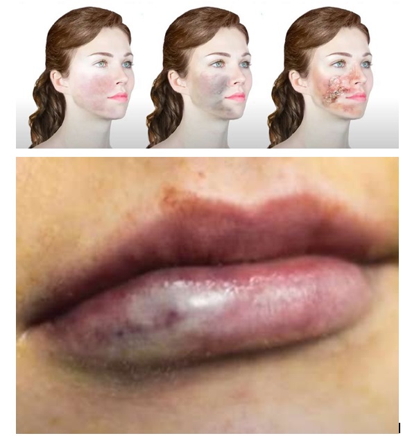
A vascular occlusion is a blockage of blood flow, ischemia is insufficient blood supply, and necrosis is irreversible tissue death. These terms describe a progression but are not interchangeable. The opportunity to prevent tissue loss depends on prompt recognition and appropriate professional treatment.
Signs that should immediately change the clinical plan
Possible warning features include unusually intense pain, sudden blanching, pale or grey skin, livedoid or reticular mottling, cool tissue, altered capillary refill, new discoloration along a vascular territory and progressive tenderness after the injection. The absence of one classic sign does not exclude injury. Local anesthetic may alter pain perception, and superficial examination can miss deeper compromised tissue.
What is capillary refill time (CRT), and what are its limitations?
CRT is assessed by briefly compressing a region of skin and observing the return of color. It can support clinical assessment of perfusion, especially when compared with adjacent areas, but it is influenced by lighting, temperature, skin characteristics and the exact tissue involved. A reassuring skin CRT does not prove that the nasal septum, deeper fat or other nearby structure is adequately perfused.
Why aspiration does not guarantee safety
Aspiration (drawing back on the syringe before injection) is sometimes taught as a way to identify an intravascular needle position. A positive blood return is important, but a negative result does not confirm safety. Contemporary reviews note false-negative aspiration due to filler viscosity, needle or cannula characteristics, test duration and other technical variables. It is unsafe to claim that aspiration ensures an injection is outside an artery. More comprehensive precautions involve anatomical expertise, controlled delivery and active monitoring, and no technique makes vascular injury impossible.[3,7]
What about hyaluronidase for threatened skin?
For suspected hyaluronic-acid-related occlusion affecting the skin, published clinical guidelines support urgent assessment and treatment with hyaluronidase by trained medical professionals, using a locally approved protocol and emergency preparedness. Treatment depends materially on the filler involved: hyaluronidase cannot be assumed to dissolve calcium hydroxyapatite, PLLA or other non-HA materials. Exact dose, preparation, injection approach and repeat assessment are clinical protocol decisions, not a universal recipe for patients to follow.[3]
Follow-up is not optional
Even when the skin initially appears improved, serial evaluation is required for evolving tissue damage, infection, wound breakdown, scarring and psychosocial consequences. Photographing the affected area with consent, recording perfusion assessments and documenting the event supports continuity of care and accurate escalation.
10. Pressure-related nasal injury: why pain and redness matter even with normal CRT
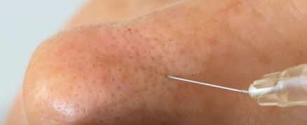
Vascular occlusion is distinct from pressure-related tissue compromise, which is especially relevant to the nasal tip and lower nose. In a confined compartment, increasing tissue pressure, hematoma formation or disruption of delicate tissue planes may endanger local structures. There may be pain, persistent redness and tenderness without the dramatic blanching often associated with intravascular filler embolization.
Why the nasal tip is different
Nasal soft tissues vary in thickness, mobility, fibrosis and prior surgical history. The limited room for expansion at the lower nose can make overfilling especially concerning. Older teaching warns against large-volume injections and quotes particular volume thresholds, but a number should not be turned into a universal safe or unsafe cutoff for every nose. Risk depends on the actual patient's anatomy, product, site, pressure and tissue response. No numerical volume guarantees safety.
Does preserved superficial capillary refill exclude pressure injury?
No. An abnormality beneath the skin can coexist with apparently adequate surface refill. Deep nasal pain after filler merits immediate clinical assessment even if the overlying skin is not pale, grey or mottled. The differential can include ischemia, compression, hematoma, infection and injury to the mucoperichondrium.
A clinically safer message for patients
Persistent, escalating or disproportionate nasal pain after non-surgical rhinoplasty should never be dismissed simply because a quick skin test looks normal. Contact the treating clinic urgently, or seek emergency assessment when severe symptoms occur. Avoid pressing repeatedly on the nose, attempting self-drainage or seeking additional filler to "correct" a painful result.
11. Case discussion: necrotic injury despite normal superficial capillary refill
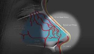
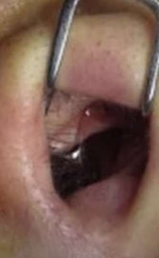
One educational case describes a patient developing serious nasal pain after non-surgical rhinoplasty. Clinicians initially considered the apparently normal skin capillary refill reassuring. Further evaluation suggested injury deeper in the nasal septum, possibly involving mucoperichondrial separation or impaired blood supply. The key lesson is not that every painful nose will become necrotic. It is that a single reassuring superficial sign cannot overrule a worrisome history.
Five diagnostic questions drawn from the case
- Exactly what was performed? Document date, product, site, amount, instrument and injection depth as accurately as available. Missing information complicates differential diagnosis and referral.
- Is there evidence of a hematoma? A localized collection may affect pressure and perfusion. An expanding or deep collection requires careful assessment.
- Could there be direct damage to septal tissue? Tissue planes around cartilage and mucoperichondrium are vulnerable to inappropriate penetration or dissection.
- Could an arterial event explain the presentation? Consider nasal and upper-lip vascular connections rather than limiting concern to a single visible skin area.
- Is the injury deeper than the skin examination can reveal? A patient with substantial pain may require specialist examination and imaging despite a normal-looking skin surface.
These are diagnostic discussion points, not a procedural guide for nasal injection. Causation in an individual case cannot be established without appropriate clinical documentation and investigation.
12. Stroke after facial filler injection
A stroke is a brain injury caused by interruption of cerebral blood supply or bleeding. In a rare mechanism, filler reaches a cerebral artery through vascular connections from facial or orbital territories. Although uncommon, embolic stroke associated with filler is a recognized serious adverse event.[1,4]
Stroke signs: remember FAST, and do not ignore other symptoms
- F, Face: new drooping or unequal facial movement.
- A, Arms: weakness or inability to lift one arm normally.
- S, Speech: slurred, unusual or difficult speech.
- T, Time: activate emergency medical services immediately.
Other warning symptoms include sudden severe headache, loss of balance, vision change, confusion or unexpected numbness. Even transient symptoms require urgent assessment. Do not drive yourself for emergency assessment if stroke is suspected.
Why stroke and blindness should be considered together
The ophthalmic and cerebral circulation have anatomical connections, so a patient with acute visual symptoms after filler may also need a neurologic assessment. UK multidisciplinary guidance underscores prompt emergency department transfer because ocular and central nervous system injury may coexist.[4]
Clinical handover should be precise
Communicate the onset time; filler manufacturer, formulation and batch when available; exact injection areas; immediately observed symptoms; any performed interventions; relevant medication and allergy history; and the patient's current neurologic and visual status. Effective handover matters more than preparing a perfect photograph before transfer.
13. Prevention begins before the syringe is opened
A safer practice starts with truthful screening, an appropriate indication and knowledge of the products' limitations. Ask about previous injections and permanent fillers, active infection, prior inflammatory responses, medications, anticoagulants, allergies, dental events, prior surgery and relevant systemic illness. The history must be interpreted rather than treated as a box-ticking exercise.
Informed consent should mention the rare serious complications
Patients should understand the realistic purpose of treatment, alternatives including not undergoing the procedure, expected swelling or bruising, delayed nodules and rare but catastrophic vascular events. A signature alone is not proof that a risk was meaningfully understood. A written aftercare sheet should explain both ordinary recovery and emergency warning signs.
Product and injector selection
Use traceable, appropriately sourced products with identifiable batch details. The suitability of a product, brand or technique should be assessed under the applicable regulator's labeling and local rules, and there is no universal FDA approval for every filler at every site. The FDA specifically recommends against some unapproved uses, including certain nasal and periorbital injections. In the UAE, clinicians must also follow the relevant local licensing, facility and device requirements.[1]
Sterile technique, training and clear emergency pathways
Aseptic preparation, appropriate equipment, anatomy training, calibrated dosing and a documented local escalation protocol are parts of prevention. Every clinic performing fillers should know its closest appropriate emergency service and how to escalate suspected vascular injury. Staff should know which symptoms warrant immediate interruption of treatment and how to communicate with the patient.
Imaging can help, but does not remove clinical responsibility
The 2025 ultrasound consensus recommends trained use of high-frequency imaging for prior filler recognition, complicated cases and selected high-risk regions. Ultrasound helps visualize anatomy and product location, but it is operator-dependent. The publication is a consensus framework, not evidence that a scanner eliminates embolic risk.[5]
14. Facial-area risk comparison: lips, cheeks, chin, nose and tear troughs
| Facial area | Common concerns | Less common but important considerations | Main safety question |
|---|---|---|---|
| Lips | Swelling, bruising, uneven distribution | Infection, delayed nodules, vascular compromise | Is pain or color change normal for this patient? |
| Cheeks and midface | Edema, contour asymmetry, malar swelling | Vascular injury, infection, delayed inflammation | Is there any unexplained worsening or asymmetry? |
| Chin and jawline | Palpability, bruising, uneven contour | Salivary gland or nerve injury, vascular injury | Are gland and nerve symptoms present? |
| Nose | Persistent contour issue, tenderness | Pressure injury, ischemia, necrosis, blindness | Is there persistent or severe nasal pain? |
| Tear trough / periorbital | Prolonged edema, visibility or Tyndall effect | Orbital vascular injury and vision loss | Has vision or eye function changed? |
| Forehead / glabella | Contour irregularity | Ophthalmic vascular connection | Is the procedure indicated, labeled and appropriately skilled? |
This matrix is educational, not a numerical ranking of event probability. Different studies and injectables have different complication reporting methods, so a single global risk score by site would be misleading.
15. Dermal filler consultation and follow-up in Abu Dhabi
Patients seeking lip filler, nose filler, chin filler, cheek filler or tear-trough treatment in Abu Dhabi should be offered more than a cosmetic price quote. A responsible consultation addresses suitability, existing filler and previous complications, individualized anatomy, alternatives, product labeling, informed consent and the clinic's arrangements for same-day assessment if a serious adverse event develops.
What to ask during a filler safety consultation
Ask which material is planned, why it is suitable, whether the intended site is covered by the relevant product labeling and local rules, how delayed complications are assessed, whether an ultrasound pathway is available where appropriate, and who will examine you if you call after hours. Ask whether the practitioner can explain, in plain language, the signs of vascular compromise and what the clinic would do immediately.
A note on marketing claims
Phrases such as "zero-risk filler," "guaranteed safe nose filler," "no chance of blindness" and "a negative aspiration makes injection completely safe" do not reflect the evidence. Equally, not every post-filler lump requires immediate dissolution or represents a dangerous infection. Helpful clinical content explains uncertainty and supports appropriate action, rather than exploiting fear or promising impossible certainty.
When is ultrasound useful in a clinic?
A qualified clinician may use ultrasound to identify previous filler, determine filler location, differentiate a lesion from an abscess or assess selected vascular problems. It should be part of an established clinical service, with appropriate equipment, training, image documentation, hygiene and pathways for escalation.[5]
16. What to expect after filler, and what not to ignore
Mild tenderness, limited edema and a small bruise can appear soon after treatment. The duration varies by location, product, technique and patient, and it is not possible to specify one recovery timeline for every filler. An individual clinician's written instructions are preferable to generic advice, especially when the patient has a history of complications.
Contact a medical professional if the skin becomes increasingly red or hot, if tenderness worsens rather than settles, if there is a new lump weeks later, or if discomfort is interfering with eating or facial movement. Seek emergency assessment immediately for changes in sight, sudden neurological symptoms, severe unexpected pain, or pale, grey, blue or mottled skin.
Dissolving filler is not a universal reset button
Hyaluronidase can break down some hyaluronic-acid products when used appropriately. It cannot be assumed to work for every brand or type of dermal filler and does not automatically reverse established tissue injury. Additional injections themselves have risks and should be performed according to a qualified clinician's assessment.[1,3]
17. What the original 2023 guide teaches, and what newer evidence changes
This article builds on Dr Tim Pearce's 2023 educational guide, which is not a peer-reviewed clinical trial or a substitute for a current emergency protocol. Its most valuable enduring contribution is the structured recognition of twelve scenarios: non-inflammatory lesions, lip lumps, inflammatory lesions, hematoma, bacterial infection, nerve injury, salivary gland injury, blindness, impending necrosis, pressure necrosis, a deeper nasal injury with normal CRT, and stroke.
The guide also emphasizes psychosocial impact, anatomical familiarity, patient selection, appropriate documentation and clear follow-up, all of which remain central to quality care. The update here is to separate those teaching themes from independently cited newer regulatory information and consensus statements, especially where the older source offers a simplified causal claim or a potentially misleading absolute.
| Clinical point | 2023 source framing | Evidence-focused 2026 interpretation |
|---|---|---|
| Aspiration | Can help avoid an intravascular injection | Positive return is informative; negative aspiration does not prove safety[7] |
| Normal CRT | May reassure in some surface assessments | Does not rule out deeper nasal or septal injury |
| Vision-loss response | Learn prevention and management | Immediate ED transfer and specialist care are paramount[4] |
| Lip lump massage | May help certain uncomplicated lumps | Use only after assessment; inappropriate for suspected ischemia or infection |
| Delayed nodules | Product, foreign-body or inflammatory reactions | Consider infection, product characteristics, host immunity and ultrasound[5,6] |
| Filler reversal | Emergency protocols exist for HA materials | Requires clinician assessment; not all fillers are dissolvable with hyaluronidase[1,3] |
| Nose volume | Higher placement volumes can increase pressure concerns | No single numeric threshold guarantees nasal safety |
18. Patient safety checklist before a dermal filler appointment
- Confirm that the clinician and facility are properly licensed for the intended procedure and location.
- Discuss the exact filler type and the realistic alternative of not having treatment.
- Disclose previous fillers, surgery, infections, medicines, allergies and prior adverse reactions.
- Ask what happens if there is severe pain, color change, vision loss or neurological symptoms.
- Ensure that the emergency referral destination and after-hours contact are clear.
- Request written aftercare, documentation of the filler used and a follow-up pathway.
- Do not accept "risk-free" or "aspiration guarantees safety" claims.
This checklist supports a conversation. It is not a certification that any particular clinic or practitioner is safe.
Frequently asked questions about dermal filler complications
1. What is the most common side effect of dermal fillers?
Bruising, tenderness and swelling are common short-term concerns, although their frequency varies by filler product, treated region and study. A mild bruise is not the same as an arterial occlusion, but symptoms must be interpreted in context.[1]
2. Are fillers safe when injected by an experienced doctor?
An appropriately trained, licensed clinician using a legitimate product can reduce preventable risks, but no person can honestly promise zero complications. Good clinical practice includes consent, individualized selection, sterile procedures and an emergency plan.
3. Can lip filler cause blindness?
Vision loss has been reported after facial filler injections due to arterial embolization. It is rare, but a new visual symptom during or after an injection requires immediate emergency care regardless of injection location.[1,4]
4. What is a vascular occlusion after filler?
It is obstruction of blood flow by filler or, in some cases, related mechanisms. It can deprive skin or other tissues of oxygen, potentially progressing to tissue injury and necrosis. A trained clinician must evaluate it urgently.[3]
5. What does filler-related vascular occlusion look like?
Warning features may include blanching, mottling, unusual skin color, severe pain and altered tissue perfusion. Presentation is variable and no single sign is reliable in isolation. Sudden visual or neurologic symptoms are emergencies.[1,3]
6. Can vascular occlusion be painless?
Absence of severe pain does not rule out ischemia, particularly when anesthesia, individual pain responses or deeper tissue injury influence symptoms. A suspicious change still requires urgent assessment.
7. What should I do if my skin turns white after filler?
If skin blanching or a pale, grey, blue or mottled area develops during or after injection, contact the treating clinician immediately and seek urgent medical attention. Do not self-inject, massage aggressively or wait for the next routine appointment.[1]
8. Can dermal filler cause skin necrosis?
Yes. Vascular obstruction or severe pressure-related injury can compromise tissue blood supply. Early diagnosis and professional emergency management are important to limit harm.[1,3]
9. Can a patient have normal capillary refill and still have necrosis?
Yes. In a described concerning nasal case, superficial skin refill was not sufficient to exclude deeper septal injury. CRT is one clinical observation, not a guarantee of tissue health.
10. Is non-surgical nose filler riskier than standard lip filler?
The anatomical consequences differ, and nasal injections have particularly significant vascular and pressure-related concerns. Risk cannot be responsibly summarized by one universal percentage for every product, practitioner and patient. Check labeling and local requirements.[1]
11. Is nose filler approved by the FDA?
The FDA lists injection of fillers into the nose among uses it does not approve for dermal fillers in the United States. Approval status in other countries is a separate regulatory question, and no statement about local authorization should be assumed from U.S. rules.[1]
12. Can aspiration prevent blindness or vascular occlusion?
No technique guarantees prevention. A positive aspiration may indicate vessel entry, but a negative aspiration can be a false negative and should never be treated as proof of safety.[7]
13. Is a cannula always safer than a needle?
Neither instrument eliminates the possibility of vascular, glandular or other injury. Risk depends on its characteristics, anatomy, placement, injection behavior, product and clinician competence.
14. What is hyaluronidase used for?
It is an enzyme that can break down hyaluronic-acid filler in appropriate circumstances. Emergency protocols for suspected HA-related skin ischemia require trained clinicians, and other filler types do not necessarily respond.[3]
15. Will hyaluronidase restore vision after filler-induced blindness?
There is no guarantee of restored vision. Specialist management is time-critical, evidence for ocular rescue interventions remains limited, and any on-site measure must not postpone emergency department transfer.[4]
16. Do all lip filler lumps need to be dissolved?
No. Some early lumps are related to swelling or product distribution and may settle. Persistent, painful, enlarging or inflamed lesions should be assessed to establish the cause before treatment.
17. Can filler lumps appear one year later?
Yes. Delayed inflammatory nodules, granulomatous reactions, filler migration and infection-related problems can develop long after injection. Timing alone does not establish the diagnosis.[6]
18. How do I know if a lip filler lump is infected?
Warmth, increasing pain, redness, drainage or fever raise concern, but infection cannot be confirmed from a photograph alone. The clinician may need examination, ultrasound and relevant investigation.[6]
19. Can dermal fillers cause a bacterial infection?
Yes, although infection is generally uncommon. Cellulitis, abscess and delayed infection-related reactions are possible. Aseptic injection technique and clear aftercare reduce avoidable risks.[1,6]
20. Can fillers cause nerve damage?
Rare nerve injury may lead to altered sensation, pain or weakness. An immediate new facial droop accompanied by other stroke symptoms must be treated as a possible neurologic emergency, not assumed to be injection-related local nerve irritation.
21. Can jawline filler injure a salivary gland?
Injury near the parotid and submandibular structures is possible. Persistent swelling or gland-region symptoms deserve evaluation and sometimes imaging or specialist referral.
22. Is under-eye filler safe for everyone?
No. The area has complex anatomy and may be unsuitable because of edema, existing anatomy, vascular risks or previous products. Suitability requires a qualified examination, an honest discussion of risks and awareness of product labeling.[1]
23. How long does bruising last after filler?
It varies. Many injection-related bruises gradually improve over days to weeks. An expanding painful hematoma, vision changes or other worrying symptoms should not be explained away as a normal bruise.
24. Can dermal filler complications occur months later?
Yes. Late-onset inflammation, lumps and other reactions are recognized. The 2024 expert consensus reviews several potential triggers and emphasizes individualized assessment rather than one universal explanation.[6]
25. Can ultrasound identify old dermal filler?
It can often help detect and localize filler material, characterize certain complications and guide evaluation when performed by an appropriately trained professional with suitable equipment.[5]
26. Can ultrasound guarantee a risk-free injection?
No. Imaging is an adjunct that may improve anatomical understanding and assessment, but it cannot remove every procedural risk. Its reliability depends on technology, training and interpretation.[5]
27. What should patients ask before getting nose or tear-trough filler?
Ask about the purpose, alternatives, site-specific risks, exact filler product, applicable regulatory labeling, previous filler identification and the clinic's plan for treating an urgent ischemic or visual event. Be wary of absolute safety assurances.
28. What happens if someone has blurred vision immediately after filler?
It requires immediate emergency assessment. Stop the injection, activate an emergency medical response and arrange urgent transfer. Do not wait to determine whether vision will recover spontaneously.[1,4]
29. What are the signs of stroke after filler?
Sudden facial drooping, arm weakness, speech difficulty, confusion, balance problems, vision changes or severe unexplained headache are warning signs. Activate emergency medical services immediately.[1,4]
30. Can fillers be dissolved at home?
No. Management depends on the material and clinical findings, and attempts at unsupervised injection or self-treatment can cause additional harm. Seek assessment from a qualified clinician.
31. Are all dermal fillers made of hyaluronic acid?
No. Fillers can contain hyaluronic acid, calcium hydroxyapatite, poly-L-lactic acid or other materials with different biological and management properties. Patients should keep records of the exact product received.[1]
32. What is the best clinic for dermal filler complications in Abu Dhabi?
The relevant question is which licensed medical service can assess the specific urgency. Suspected eye or stroke events need immediate hospital emergency care, while stable cosmetic lumps or delayed nodules may be evaluated by an experienced, appropriately licensed aesthetic specialist. Avoid delaying urgent treatment while searching for a preferred clinic.