

Facial Bone Aging in Abu Dhabi : How the Orbit, Midface and Jaw Change with Age
Why facial rejuvenation must treat structure, not just skin.
Aging is not only a skin problem. In a 3D CT study of 120 Caucasian adults, the bony orbit enlarged with age, the glabellar and maxillary angles became more acute, the pyriform aperture became larger, and the mandible became shorter and lower while its angle opened. These skeletal changes reduce the support beneath the soft-tissue envelope and can contribute to under-eye hollows, brow and lid changes, deeper nasolabial folds, reduced nasal support, weaker chin projection, prejowl hollowing and a less defined jawline. Facial rejuvenation should therefore begin with structural diagnosis: skin quality, fat distribution and bone support can each cause the same visible sign of aging, and each may need a different strategy.
Why This Study Changed the Way We Think About Facial Aging
For decades, facial aging was described mainly as skin laxity, gravity and soft-tissue descent. Shaw and colleagues expanded that model by examining the facial skeleton itself. Their premise was simple but clinically important: if the bony framework changes, the soft tissue draped over it will project differently.
The study combined upper-face, midface and lower-face measurements in one comparatively large 3D CT dataset, treating the face as a connected structural system. This matters because the same complaint can arise from different layers. A tear trough may reflect soft-tissue change, orbital rim remodeling, or both. A deeper nasolabial fold may involve skin, fat, muscle and reduced maxillary support. A weak jawline may reflect skin laxity, subcutaneous tissue, platysma, mandibular shape or several together.
Study Design : Who Was Included and How the Face Was Measured
The investigators analyzed facial-bone CT scans from 120 Caucasian subjects, 60 women and 60 men. Per sex, 20 subjects were in a young group (20–40 years), 20 in a middle group (41–64) and 20 in an older group (65 or above). CT slices were 1.0 mm thick, each scan was reconstructed in three dimensions, and scans were standardized to the sella-nasion cranial base. Edentulous patients were deliberately excluded, because tooth loss can independently alter mandibular anatomy and confound age-related change.
The subjects were imaged for clinical reasons such as possible facial fracture, severe sinusitis or soft-tissue disease, and only scans without fracture or bony involvement were included. This was a cross-sectional comparison of different people at different ages, not a longitudinal study of the same people over decades.
What was measured?
| Region | Measurements |
|---|---|
| Orbit | Orbital aperture width, orbital aperture area, and curvilinear distances to the superior and inferior orbital rims at nine standardized positions |
| Midface | Glabellar angle, maxillary angle, pyriform angle, pyriform aperture area |
| Mandible | Bigonial width, ramus breadth, ramus height, mandibular body height, mandibular body length, mandibular angle |
The Orbit Gets Larger with Age
The orbital aperture widened and increased in area in both sexes. Male orbital width rose from a mean of 38.7 mm in the young group to 40.5 mm in the old group; in women the means were 35.7 mm and 38.3 mm. Orbital area rose from 1072 to 1191 mm² in men and from 930 to 1106 mm² in women. The change was not uniform around the rim: the superomedial and inferolateral portions remodeled most.
Timing differed by sex. Several orbital changes were statistically evident between young and middle-aged women, while comparable changes in men were more prominent between middle and older age. That is a group-level pattern in this sample, not proof that every woman ages earlier than every man.
Why orbital enlargement matters
The bony orbit is the fixed frame around the eye. When it enlarges and specific rim segments recede, the relationship between bone, orbital contents, eyelids and brow changes, so a purely skin-based explanation of periorbital aging can miss a structural component.
How Orbital Bone Remodeling Can Change the Upper Face and Under-Eye Area
The authors propose that superomedial rim remodeling can unmask medial upper-lid fat, an appearance often attributed only to a weakening orbital septum. A more acute glabellar angle may contribute to medial brow descent and glabellar creases. Inferolateral remodeling and increased orbital width may contribute to crow's-feet, lower-lid lag and lateral orbital changes. In the lower orbit, soft tissue can roll over a more recessed bony ledge, adding to lower-lid fat prominence and a deeper nasojugal groove.
This is not a claim that bone is the sole cause of every under-eye hollow or brow change. It is a structural working hypothesis: bony change alters the platform under the soft-tissue envelope, and the visible pattern emerges from the interaction of bone, fat, septum, muscle and skin.
The Orbital Rim Does Not Age as One Uniform Circle
The curvilinear measurements show the enlargement was regionally patterned. In men, significant superior-rim differences were concentrated at medial positions (x coordinates 10–40), while inferior-rim differences were more lateral (x 50–90). In women, superior-rim change extended from x 10–60 and inferior-rim change was strongest from x 60–90.
This helps explain why periorbital aging is not symmetrical around the eye. Medial upper-lid change, lateral hooding, lower-lid support and the tear-trough region can evolve differently. "Under-eye aging" is too broad a diagnosis on its own: medial hollowing, the lateral lid-cheek transition, brow support and lower-lid laxity can be different anatomic problems, even in the same patient.
The Midface Also Loses Skeletal Projection
Two key midface findings were progressive decreases in the glabellar and maxillary angles. The maxillary angle fell from 62.2° to 53.3° in men and from 64.9° to 54.0° in women (young to old). The glabellar angle fell from 72.3° to 67.6° in men and from 76.8° to 71.7° in women. The pyriform angle did not change significantly, but pyriform aperture area increased from 624.5 to 663.4 mm² in men and from 486.4 to 555.4 mm² in women. The authors interpret this as an age-related reduction in midface skeletal support.
Why maxillary remodeling can deepen the nasolabial fold
The maxilla supports the central midface. When its projection decreases, the malar fat pad and surrounding soft tissue sit on a less projecting platform, which can make the nasolabial crease look more prominent. Injecting only the visible fold may therefore miss the true anatomy, because correction focused on the crease can ignore the platform that normally supports the cheek and perioral tissue.
The source also links a larger pyriform aperture to reduced nasal support and projection. This is a structural contribution, not a claim that every aging nose needs volume augmentation.
The Mandible Becomes Shorter, Lower and More Open
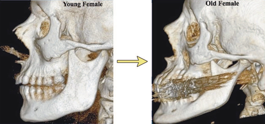
The mandible showed some of the clearest changes. Body length fell from 90.4 to 83.0 mm in men and from 86.9 to 80.8 mm in women. Body height fell from 35.4 to 32.3 mm in men and from 31.5 to 27.7 mm in women. Ramus height also fell in older subjects, while the mandibular angle rose from 112.3° to 125.6° in men and from 119.9° to 132.0° in women. Bigonial width and ramus breadth did not change significantly, which shows that skeletal aging is patterned remodeling rather than uniform shrinking.
Summary of key measurements (young to old group means)
| Measurement | Men | Women | Direction |
|---|---|---|---|
| Orbital width | 38.7 → 40.5 mm | 35.7 → 38.3 mm | Increases |
| Orbital area | 1072 → 1191 mm² | 930 → 1106 mm² | Increases |
| Glabellar angle | 72.3° → 67.6° | 76.8° → 71.7° | More acute |
| Maxillary angle | 62.2° → 53.3° | 64.9° → 54.0° | More acute |
| Pyriform aperture area | 624.5 → 663.4 mm² | 486.4 → 555.4 mm² | Increases |
| Mandibular body length | 90.4 → 83.0 mm | 86.9 → 80.8 mm | Decreases |
| Mandibular body height | 35.4 → 32.3 mm | 31.5 → 27.7 mm | Decreases |
| Mandibular angle | 112.3° → 125.6° | 119.9° → 132.0° | More open |
| Bigonial width / ramus breadth | No significant change | Stable | |
A wider mandibular angle combined with loss of mandibular height and length can soften the crisp structural boundary of the lower face. The source proposes this skeletal change contributes to loss of jawline definition and reduced chin projection, in addition to soft-tissue and neck aging.
Why Skeletal Aging Matters for the Chin, Prejowl and Neck
The mandible is the foundation of the lower face. The authors note that a normal ramus height and body length give stronger support for soft-tissue repositioning, whereas a short ramus, open angle and short body give less. Decreased chin projection and the prejowl sulcus are specifically linked to deficient skeletal support, and mandibular volume loss can contribute to neck aging by reducing the platform beneath the platysma. Jowling and neck laxity are therefore not always purely skin problems.
Men and Women Showed Different Timing Patterns
Women often showed significant orbital and midface differences between the young and middle groups, while men more often showed them between the middle and older groups. For example, female orbital width increased significantly from young to middle age, whereas the male increase was clearest between young and old. The investigators did not claim sex alone determines facial aging, and with only 20 subjects per sex-by-age subgroup, this should not become a fixed biological clock. The useful message is that structural aging is heterogeneous, not synchronized across the face.
Bone Loss and Soft-Tissue Volume Loss Are Related but Not Identical
Facial fat can atrophy or redistribute while the orbit, maxilla and mandible change their dimensions at the same time, so a patient can look deflated for more than one reason. Replacing soft-tissue volume does not regrow bone. Strategically placed volume can camouflage a less projecting skeletal platform and recreate some lost support visually, whereas surgical skeletal augmentation changes the framework directly. A layered diagnosis asks whether the dominant deficit is skeletal projection, fat volume, skin quality, soft-tissue descent or a combination.
What the Source Says About Rejuvenation Strategies
The authors argue that soft-tissue redraping and skin reduction alone may be incomplete when the skeleton has lost projection. They discuss three broad approaches: soft-tissue fillers (nasolabial fold, glabellar crease, malar region, nasojugal groove, lips), autologous fat grafting where fat loss is a major driver, and alloplastic skeletal augmentation for selected patients with intact occlusion and a structural bony deficiency. These are surgical concepts from a 2011 plastic-surgery paper, not a one-size-fits-all algorithm.
It does not compare hyaluronic-acid filler with CaHA or PLLA, does not study Endolift, XERF, HIFU, RF microneedling or laser, and does not test whether any modern nonsurgical treatment can reverse skeletal aging. It is an anatomy-and-aging study that informs diagnosis and planning.
How This Anatomy Changes Nonsurgical Aesthetic Planning
The study's value is diagnostic rather than prescriptive. Lower-eyelid hollowing may combine orbital rim remodeling, fat-compartment change and skin quality loss. A heavy nasolabial fold may combine maxillary retrusion with soft-tissue descent. A weak jawline may combine mandibular remodeling with laxity and fat change. The practical question is not "Which product should be injected?" but "Which layer is creating the visible problem?" Two patients of the same age can need completely different plans, because chronologic age does not identify the dominant deficit.
The 2011 study predates today's device combinations. Injectable volume can improve contour over a deficient platform, but placement and amount depend on anatomy. Collagen stimulators may improve soft-tissue quality, and tightening devices can improve selected laxity, but this paper does not show that either reverses orbital, maxillary or mandibular remodeling. Treatment should start with the visible deficit and the layer that produces it: a patient with good projection and mild laxity is a different candidate from one with major midface retrusion, even if both just say "sagging."
Structural Diagnosis Matrix : What Might Be Under the Visible Complaint?
| Visible concern | Possible structural contribution | Other layers that still need assessment |
|---|---|---|
| Under-eye hollow / nasojugal groove | Inferior orbital rim recession and enlarged orbital aperture | Fat compartments, septum, skin quality, edema, lower-lid laxity |
| Medial brow descent / upper-lid hollowing | Superomedial orbital rim remodeling and more acute glabellar angle | Frontalis/orbicularis balance, upper-lid fat, dermatochalasis |
| Prominent nasolabial fold | Reduced maxillary projection and malar soft-tissue support | Cheek fat loss, ligamentous support, skin, muscle activity |
| Reduced nasal support | Larger pyriform aperture | Cartilage, skin, tip support, prior surgery |
| Weakening chin projection | Shorter, lower mandible and altered geometry | Soft-tissue thickness, dental occlusion, baseline chin anatomy |
| Prejowl hollow / jowling | Reduced mandibular skeletal support | Fat distribution, retaining structures, platysma, skin laxity |
| Blurred jawline / heavy neck | More open mandibular angle, reduced lower-face support | Subcutaneous fat, platysma, skin excess, hyoid/neck anatomy |
A More Precise Way to Discuss "Volume Loss"
"The face loses volume" hides several events: fat atrophy, change in the bony platform, and descent or redistribution of tissue. A treatment that hydrates skin may improve texture without restoring projection. A filler restores contour without tightening excess skin. A tightening procedure reduces selected laxity without recreating a deficient maxilla. Surgical repositioning moves tissue but may still need volume in a structurally deflated face. The most natural result comes from matching each intervention to the layer that is actually deficient.
Why "just tighten the skin" can miss the problem
If the skeleton is less projecting while soft tissue also loses volume and elasticity, tightening the surface cannot recreate every lost three-dimensional relationship. Equally, adding volume without understanding where support has changed can create heaviness rather than restoration. Natural rejuvenation rests on proportion, anatomy and the interaction of layers, not on treatment trends.
Facial Bone Aging Is Not the Same in Every Person
The paper shows population-level trends, but baseline skeletal anatomy, sex, dentition, genetics, soft-tissue distribution and environmental exposure all influence how remodeling becomes visible. The numbers are group means, not diagnostic thresholds: an orbital width or mandibular angle beyond a given value does not by itself mean a patient is "aged" or needs treatment. The study did not use soft-tissue segmentation, standardized aesthetic grading or patient-reported scores to link each millimeter of bone change to a visual effect. For an individual, photographs, animation, palpation, dental context, soft-tissue thickness and the face-neck relationship all change the interpretation.
Study Strengths and Limitations
Strengths: 120 subjects balanced by sex and across three age strata; direct 3D CT measurement instead of surface photography; orbit, midface and mandible examined together using linear, angular and area measurements; exclusion of edentulous patients; and quantitative statistical testing.
Limitations:
- Cross-sectional design: different people in each age group, so findings are associations, not observed individual change.
- All participants were Caucasian, so results should not be generalized to every population without further evidence.
- Scans were obtained for clinical indications, not from a prospectively recruited healthy cosmetic cohort.
- Aesthetic consequences were inferred; wrinkle severity, tear-trough depth, nasolabial fold grade, chin projection and jawline attractiveness were not measured as paired outcomes.
- Published in 2011: the anatomy remains relevant, but products, implants and technologies have evolved.
Evidence integrity: the strongest conclusion is that the facial skeleton remodels with age and gives progressively different support to overlying tissue. The study supports structural diagnosis; it does not prove that any filler, implant, energy device or protocol is superior.
A Practical Structural Assessment for Facial Aging in Abu Dhabi
For patients in Abu Dhabi considering facial rejuvenation, it helps to split the face into structural questions. Around the eyes, is the main issue muscle activity, skin quality, orbital support, fat prominence or hollowing? In the midface, is there lost projection, fat deflation or mainly skin laxity? In the lower face, is the weak contour from chin projection, mandibular support, jowling, platysma, fat or all of these? This avoids over-treating one layer while ignoring another, and it keeps expectations realistic: improving collagen or skin tightness is not the same as restoring skeletal projection, and neither equals soft-tissue repositioning.
Facial Aging Consultation with Dr. Erfan Rahmani in Abu Dhabi
An evidence-based consultation should start with anatomy rather than a product name. Dr. Erfan Rahmani can assess orbital support, midface projection, chin and jawline structure, fat distribution, skin laxity and previous treatments before discussing the most appropriate nonsurgical options, or whether surgical evaluation would better match the degree of structural aging. This page does not suggest that every age-related skeletal change should be injected or augmented; it explains why rejuvenation becomes more precise when the underlying architecture is understood.
More information : www.drerfanrahmani.com
Frequently Asked Questions About Facial Bone Aging
Does the facial skeleton really change with age?
Yes. In this study, the bony orbit, midface and mandible all showed measurable age-associated changes.
What happens to the orbit with age?
Orbital aperture width and area increased, with prominent remodeling of the superomedial and inferolateral rims.
Can orbital bone aging contribute to under-eye hollows?
The authors propose that rim recession can change soft-tissue support and contribute to lower-lid fat prominence and a deeper nasojugal groove.
What happens to the maxilla with age?
The maxillary angle decreased significantly in both sexes, consistent with reduced midface skeletal projection.
Does the pyriform aperture change?
Yes. Its area increased significantly with age, while the pyriform angle did not change significantly.
What happens to the jawbone with age?
Mandibular body length and height decreased, ramus height decreased, and the mandibular angle became more open.
Does the jaw simply become narrower?
Not in this study. Bigonial width and ramus breadth showed no significant age-related change.
Can jawbone aging affect the jawline?
Yes. The source proposes that a more open angle and reduced mandibular dimensions can contribute to reduced lower-face definition.
Can skeletal aging affect the neck?
The source suggests reduced mandibular support can contribute to the appearance of neck soft-tissue and platysmal laxity.
Why can the nasolabial fold deepen with age?
One contributor may be reduced maxillary support beneath the cheek and malar fat pad, in addition to soft-tissue aging.
Is facial aging only caused by bone loss?
No. The source frames aging as a combination of skeletal change, fat loss and soft-tissue and skin change.
Does this study prove fillers reverse bone aging?
No. It discusses volume replacement strategies but did not test a filler protocol against skeletal aging.
Does this study compare Sculptra, Radiesse or hyaluronic acid?
No. Those head-to-head comparisons are outside the study.
Does it study Endolift, XERF or HIFU?
No. The paper predates these treatments and focuses on skeletal anatomy and rejuvenation concepts.
Were the same patients followed as they aged?
No. It was a cross-sectional study comparing different age groups.
Were all ethnicities represented?
No. All 120 subjects were Caucasian, which limits generalizability.
Why were edentulous patients excluded?
To reduce the influence of tooth loss and related mandibular remodeling on the age comparisons.
What is the key message for aesthetic treatment planning?
Identify which anatomic layer is responsible for the visible change before selecting a treatment.
Where can I learn more in Abu Dhabi?
For consultation information with Dr. Erfan Rahmani, visit www.drerfanrahmani.com