
Endolift 1470 nm Study : What the 60-Day Evidence Shows (and Doesn't)
In a 2023 case series of five patients with grade 1–2 facial rhytids, 1470 nm diode-laser endolifting was associated with increased dermal and subcutaneous thickness on ultrasound at 60 days. The authors also reported visible malar and mandibular improvement, and all participants reported some level of satisfaction. The study was preliminary, uncontrolled and too small to predict the result or complication rate for an individual patient.
If you are researching Endolift 1470 nm in Abu Dhabi, this guide explains what the published study measured, what it found and where its limits are.
Facial Ageing Is a Multi-Layer Process
The study describes the face as a composite of bone, muscle, aponeurotic tissue, fat and skin. Superficial fat compartments are separated by fascial tissue and retaining ligaments, so ageing is not just surface wrinkling. Volume changes occur at different rates in different compartments, which alters facial contour and proportions.
The paper also describes the superficial musculo-aponeurotic system (SMAS) as a network of collagen, elastic fibers and adipocytes connecting the facial muscles to the dermis. The dermis itself contains a three-dimensional network of collagen and elastic fibers that provides support, resistance and retraction, and these properties decline with age. The authors also describe bone resorption, loss of fat and muscle, upper-face skeletalization and increasing lower-face laxity.
What Are Facial Rhytids?
The authors distinguish three types of lines:
- Superficial wrinkles are related to skin texture.
- Mimetic lines are caused by repeated movement combined with dermal elastosis.
- Deeper folds result from overlapping skin linked to laxity, intrinsic ageing, reduced tone, skeletal atrophy, gravity and tissue descent.
This matters because a tissue-tightening procedure does not address every component of ageing equally. Dynamic lines, skeletal support, fat distribution, severe skin excess and pigment concerns each need separate assessment.
Why a 1470 nm Diode Laser?
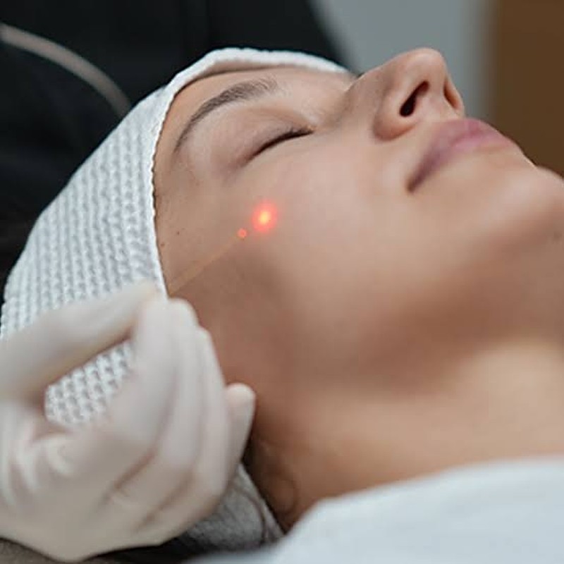
Laser light is monochromatic, coherent and collimated, and its tissue effect depends on absorption by chromophores such as water, hemoglobin, melanin and proteins. The 1470 nm wavelength is highly absorbed by water and relatively weakly absorbed by melanin and hemoglobin. It is delivered through a flexible optical fiber, and a transcutaneous guide beam helps the operator see where energy is being deposited.
The goal is not superficial resurfacing. Energy is delivered through a fiber placed in deeper tissue planes.
How Endolifting Is Proposed to Work
The authors cite earlier laser-assisted liposuction and endolifting work describing thermal effects on the dermis. These include remodeling of existing collagen, stimulation of new collagen, increased dermal thickness and skin retraction, without the large wounds of open surgery.
This study did not perform biopsies. Instead, the investigators used ultrasound and interpreted greater dermal thickness, increased hypoechogenicity and thicker trabecular patterns as compatible with collagen increase and tissue remodeling.
Who Was Included in the Study?
Participants were 40 to 75 years old, of either sex, with grade 1–2 rhytids on the modified Fitzpatrick scale. Eligible findings included dynamic and static wrinkles, marionette lines, jowl-line change, reduced definition of the maxillary or mandibular border, and localized adiposity in the zygomatic and anterior cervical areas. Laboratory tests had to be within normal limits.
The study did not test severe facial ageing, so its findings apply most directly to mild-to-moderate changes.
Exclusion Criteria
| Exclusion criterion | Why it matters when reading the results |
|---|---|
| Pregnancy or breastfeeding | These populations were not studied. |
| Uncontrolled medical disease | Medically unstable patients were not tested. |
| Botulinum toxin, PDO threads or dermal filler in the preceding 6 months | Intended to reduce confounding from recent aesthetic procedures. |
| Active disease in the treatment area | Potential safety and outcome confounder. |
| Autoimmune or collagen disease | May alter healing or tissue response. |
| Active cancer | Excluded from the study population. |
| Prolonged steroid use | Potential effect on tissue response and healing. |
| Previous radiotherapy in the treatment area | Potentially altered tissue quality. |
| Allergy to local anesthetic | Relevant because local and tumescent anesthesia were used. |
How Outcomes Were Measured
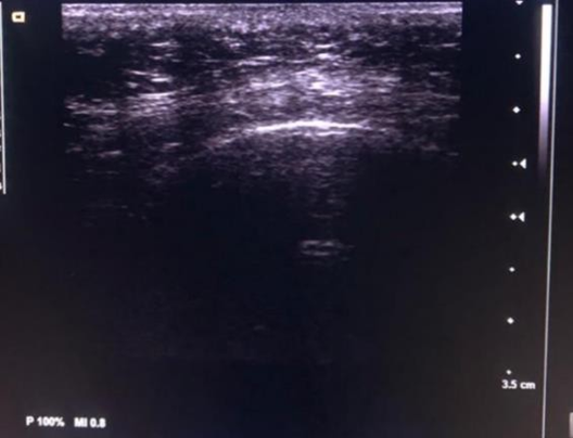
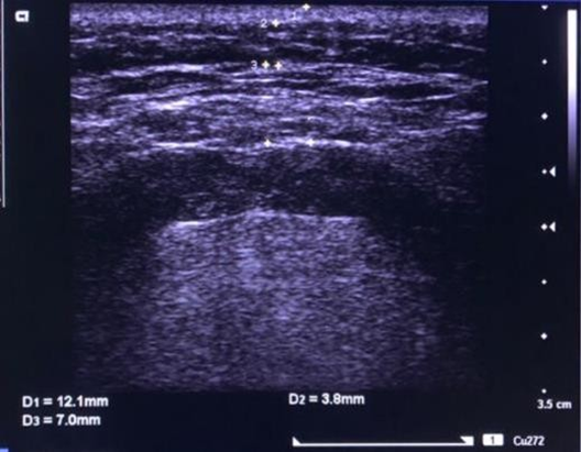
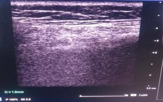
- Ultrasound : A Siemens Acuson X300 with a 7.5–12 MHz linear transducer. Longitudinal, transverse and oblique sweeps documented epidermal, dermal and subcutaneous thickness across facial, periorbital and zygomatic regions.
- Standardized photography : Frontal, 45-degree oblique and 90-degree lateral views of both hemifaces, to limit distortion from lighting, head position and camera angle.
- Patient satisfaction : A five-category Likert scale from "extremely satisfied" to "not satisfied."
All three were reassessed at 60 days.
The Treatment Protocol Used in the Study
| Protocol element | Study-specific detail |
|---|---|
| Preparation | Surgical kit; asepsis and antisepsis of face and neck with non-alcoholic bactericidal solutions; laser and optical fiber prepared |
| Marking | Vector-based treatment design |
| Entry-point anesthesia | 1% or 2% lidocaine |
| Broader anesthesia | Modified Klein tumescent anesthesia via facial infiltration cannula |
| Laser platform | Velas diode laser (GigaLaser INC, Germany / South Korea) |
| Emission mode | Continuous |
| Power (hemiface and neck) | 6 W |
| Power (lower eyelid) | 2–3 W |
| Fiber (face) | 400 µm |
| Fiber (lower eyelid) | 200 µm |
| Plane | Deep dermis and subcutaneous tissue |
| Movement | Fan-like movements along planned vectors |
| Passes | 10–15 |
Post-Procedure Care in the Study
The study's regimen included analgesics, anti-inflammatory medication, cryotherapy for 3–5 days, prophylactic antibiotics for 5–8 days, avoiding heat and steam, not lowering the head during the first 8 days, SPF 50+ sun protection and scheduled follow-up. This was one study's protocol, not universal advice. Medication, cryotherapy and activity restrictions should be individualized by the treating clinician.
Ultrasound Results at 60 Days
The authors reported increased thickness of the dermis and subcutaneous tissue, with the greatest changes in the malar and submental regions. The largest difference was 3.6 mm in the right submental region, and a smaller 0.4 mm difference was noted in the left malar region.
| Region | Selected values reported in Table 1 (mm) |
|---|---|
| Periorbital dermis | 0 to 1.8 across patients/sides |
| Malar dermis | 0.4 to 3.5 across patients/sides |
| Submental dermis | 1.4 to 3.6 across patients/sides |
| Upper-third subcutaneous tissue | 0 to 4.7 across patients/sides |
| Middle-third subcutaneous tissue | Positive and negative values; variable response |
| Lower-third subcutaneous tissue | Positive and negative values; variable response |
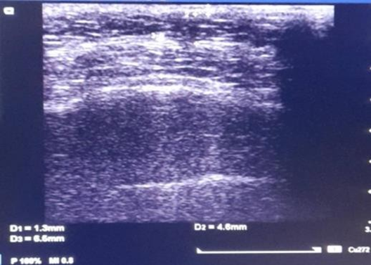
How the Authors Interpreted the Images
Before treatment, the epidermis appeared as an echogenic line with reduced refringence, and the dermis looked relatively hypoechoic. At follow-up, the authors described greater epidermal echogenicity, thicker dermis and trabeculae, and increased hypoechogenicity. The subcutaneous tissue showed more prominent undulating trabeculae between fat cells. They interpreted these changes as compatible with increased endogenous collagen and structural remodeling.
Photographic Outcomes : Day 1 vs Day 60
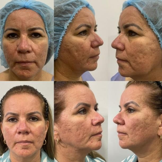
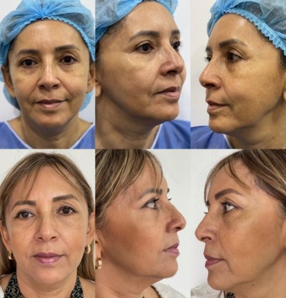
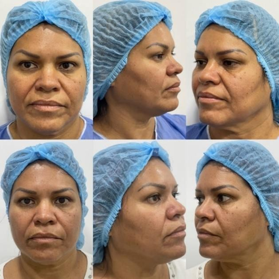
The authors reported significant photographic improvement in the malar and mandibular areas, consistent with the ultrasound findings. The paper includes three patient panels comparing day 1 with day 60 from frontal and oblique or lateral views. Actual study photographs are more informative than generic marketing images, but a five-patient series cannot establish the average outcome for a broader population.
Patient Satisfaction
| Satisfaction category | Reported result |
|---|---|
| Extremely satisfied | 40% |
| Very satisfied | 20% |
| Moderately satisfied | 40% |
| Little satisfied / not satisfied | 0% |
| Overall reporting some satisfaction | 100% of 5 patients |
This was 100% satisfaction in five patients, not a proven 100% success rate. Satisfaction is also shaped by expectations, counseling, recovery experience and baseline severity, so it is not the same as objective efficacy.
What the Authors Concluded
The authors describe 1470 nm endolifting as efficient for managing facial rhytids in this small series. They compare their observations with earlier work by Nilforoushzadeh and colleagues on 1470 nm endolifting and with biometric studies suggesting increased skin thickness, density and elasticity. This study is not a controlled replication of that work.
Most importantly, the authors call their results preliminary and not conclusive and recommend larger studies with designs that can test hypotheses more rigorously.
Complications and Recovery
No complications were reported in these five patients. That does not mean the complication risk is zero, because rare events cannot be estimated from such a small sample.
The paper calls the technique minimally invasive with rapid recovery and cites a three-to-five-day recovery context from earlier literature. The study's own aftercare involved several days of medication, cryotherapy and precautions. "Rapid recovery" is more defensible than "zero downtime," since swelling, bruising and tenderness vary between patients and areas.
Jawline, Submental Area and Lower Eyelid
Jawline and submental area : Photographic improvement was reported in the mandibular area, and the largest ultrasound change was in the submental region. The 3.6 mm figure was one tissue-thickness difference in a five-patient dataset, not a guaranteed lift or a fixed amount of fat reduction. A consultation should determine whether the problem is skin laxity, localized fat, skeletal anatomy, tissue descent or a combination.
Lower eyelid: The protocol used lower power and a smaller fiber for this area, and periorbital dermal thickness was measured. However, there was no separate large analysis of eyelid outcomes, so the study cannot support a promise of eyelid lift or reduced eye bags.
Does This Study Prove Endolift Is Better Than Surgery?
No. Patients were not randomized to Endolift versus facelift, and no comparative outcomes were measured. The two approaches address different degrees of ageing, and this evidence is most relevant to grade 1–2 rhytids. It does not establish equivalent correction for severe skin excess or major structural descent.
Strengths and Limitations of the Study
Strengths
- Clearly defined population using a modified Fitzpatrick rhytid scale
- Documented inclusion and exclusion criteria
- Three outcome types: ultrasound, standardized photography, satisfaction
- Defined 60-day follow-up
- Transparent technical details
- Actual numerical ultrasound differences published
Limitations
- Only five patients
- No randomized or untreated control group
- No described blinded assessment
- No follow-up beyond 60 days
- No validated wrinkle score per patient
- Variable ultrasound results, including negative subcutaneous values, not deeply analyzed
- Cannot separate collagen remodeling from edema resolution, contraction or architectural change
- No comparison with other tightening modalities
What This Study Supports and What It Does Not Prove
Supports
- Feasibility of 1470 nm endolifting in five patients with grade 1–2 rhytids
- Measurable ultrasound tissue changes at 60 days
- Increased dermal thickness, especially malar and submental
- Photographic malar and mandibular improvement
- Some satisfaction in all five participants
- Separate parameters for face/neck and lower eyelid
- Minimally invasive treatment with rapid recovery in this cohort
Does not prove
- A 100% success rate in the general population
- Zero risk of complications
- A guaranteed 3.6 mm improvement
- Permanent results
- Superiority over facelift, HIFU, radiofrequency or fillers
- That one set of parameters suits every device or anatomy
- Durability beyond 60 days
- Equivalent outcomes in severe laxity
Why the 60-Day Follow-Up and Grade 1–2 Boundary Matter
Repeating ultrasound and photography at 60 days reduces the risk of mistaking acute swelling for the final result. Still, 60 days is short for a procedure discussed in terms of collagen remodeling. The study does not say whether changes persisted at six months or a year, or whether retreatment was needed.
The grade 1–2 restriction is equally important. A broad claim such as "Endolift treats all facial sagging" is not supported. The study evaluated selected patients with early rhytid grades, and advanced cases were not tested.
A Practical Consultation Framework for Endolift in Abu Dhabi
- Define the dominant concern: wrinkles, lower-face laxity, jowling, jawline definition, submental fullness, lower-eyelid laxity or a combination.
- Decide whether a minimally invasive approach fits the degree of ageing or the desired correction is closer to a surgical endpoint.
- Review medical history, medications, skin disease and recent toxin, filler or thread procedures.
- Assess whether volume should be preserved, reduced or redistributed, rather than assuming everyone needs fat reduction.
- Use standardized baseline photography, and ultrasound when clinically appropriate.
- Explain that the published evidence consists of small case series and cannot be converted into guaranteed percentages.
- Discuss recovery as variable and avoid "zero downtime" wording.
- Review alternatives and combination options according to anatomy.
- Judge results over weeks to months, not immediately.
Endolift 1470 nm in Abu Dhabi with Dr. Erfan Rahmani
For patients researching Endolift 1470 nm in Abu Dhabi, the key question is whether the procedure matches your anatomy and the level of correction you expect. A less-defined jawline can come from laxity, fat distribution, volume loss, skeletal proportions or a mix of these.
Dr. Erfan Rahmani can assess facial laxity, jawline definition, submental fullness, lower-eyelid anatomy, volume distribution and previous aesthetic treatments to decide whether Endolift is appropriate. Learn more at www.drerfanrahmani.com.
Frequently Asked Questions
What did the 2023 Endolift study investigate?
It evaluated a 1470 nm diode-laser endolifting technique in five patients with grade 1–2 facial rhytids, using ultrasound, standardized photography and patient satisfaction at 60 days.
How many patients were included, and what age range?
Five patients, with inclusion criteria of 40 to 75 years.
What was the largest ultrasound change reported?
A maximum thickness difference of 3.6 mm in the submental region.
Did the study show jawline improvement?
The authors reported significant photographic improvement in the malar and mandibular areas.
Were patients satisfied?
Yes. All five reported satisfaction: 40% extremely, 20% very and 40% moderately satisfied.
Does 100% satisfaction mean Endolift always works?
No. This was a five-patient case series, so the percentage cannot be generalized.
Were there complications?
None were reported in this five-patient cohort, which does not prove the risk is zero.
How long was follow-up?
Sixty days.
What wavelength, fiber sizes, power and passes were used?
A 1470 nm diode laser with a 400 µm fiber for the face and a 200 µm fiber for the lower eyelid. Power was 6 W for the hemiface and neck and 2–3 W for the lower eyelid, with 10–15 fan-pattern passes. These are study-specific parameters, not universal settings.
Does this prove Endolift increases collagen?
No. The authors interpreted ultrasound changes as compatible with increased collagen, but the study did not quantify collagen with biopsy.
Is Endolift better than a facelift?
The study did not compare the two, so superiority cannot be concluded.
Is there zero downtime?
No. The paper describes rapid recovery and a three-to-five-day recovery context, and the study protocol included several days of aftercare.
Can Endolift treat the lower eyelid?
The study included a lower-eyelid protocol but did not provide a separate large outcome analysis for that area.
What is the main limitation of this evidence?
A sample of five patients, no control group and follow-up limited to 60 days.
Where can I learn more about Endolift in Abu Dhabi?
Visit www.drerfanrahmani.com for information and consultation details with Dr. Erfan Rahmani.