
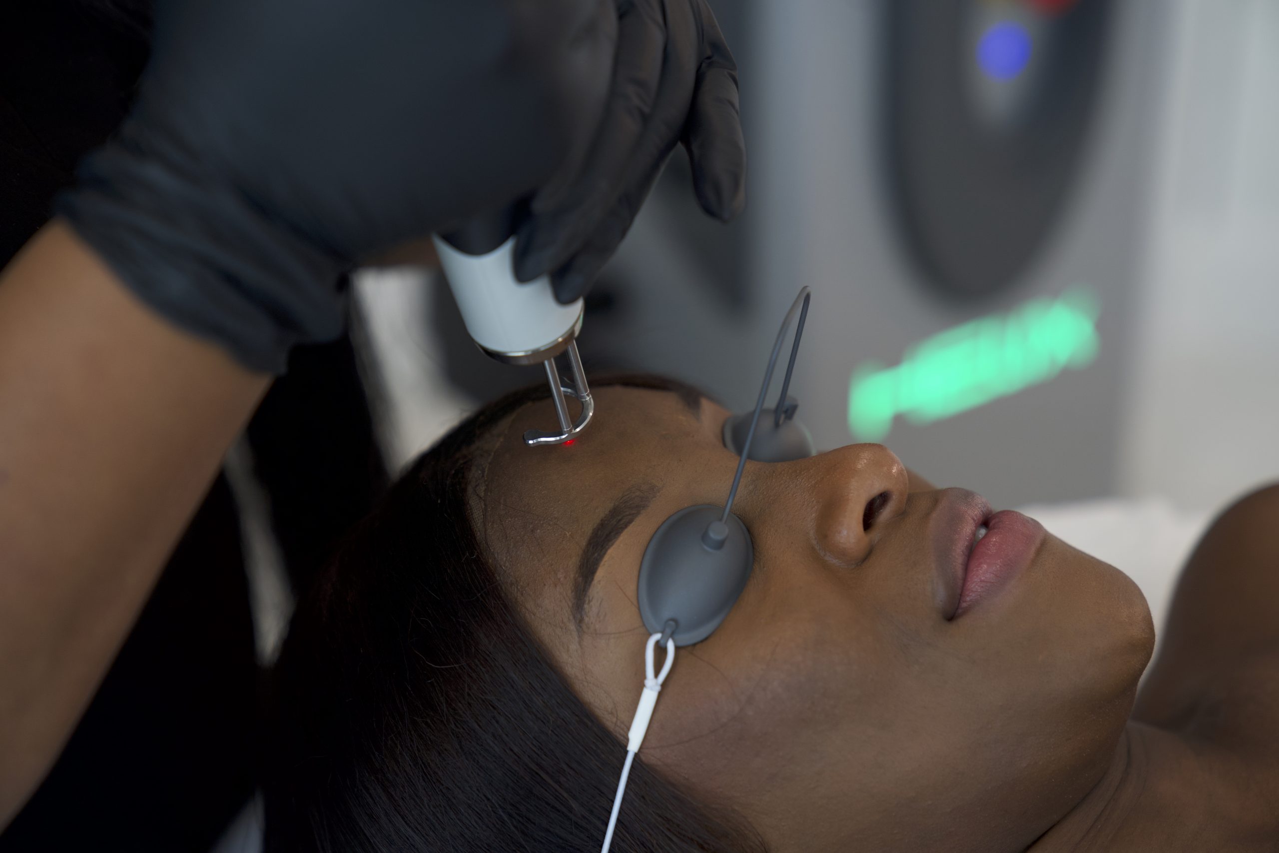
What is a picosecond laser ?
A picosecond is one trillionth of a second, or 10^-12 seconds. By comparison, a nanosecond is 10^-9 seconds. The defining difference is therefore pulse duration: picosecond systems deliver laser energy over a substantially shorter period than traditional Q-switched nanosecond systems.
The source review explains why this matters. When a similar amount of energy is delivered over a shorter pulse, peak power rises. This can increase photomechanical and photoacoustic effects on pigment particles while reducing reliance on prolonged heating of surrounding tissue.
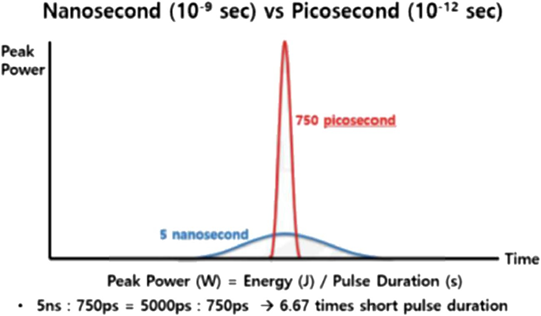
Photoacoustic effect, thermal relaxation time, and stress confinement
The review distinguishes two important physical concepts. Thermal relaxation time describes how long a target takes to lose approximately half of the heat generated during laser exposure. Selective photothermolysis is favored when the laser pulse is shorter than the thermal diffusion time of the target.
Picosecond technology adds another important concept: stress relaxation time. When tissue rapidly expands after energy absorption, acoustic shock waves are generated. A pulse shorter than the acoustic diffusion or stress-relaxation time favors selective photoacoustic disruption. This is especially relevant to tattoo ink particles, where fragmentation rather than bulk heating is a central treatment mechanism.
The authors describe picosecond treatment as being more closely associated with stress confinement, while nanosecond treatment is more closely associated with thermal confinement. The practical implication is not that heat disappears, but that the balance of physical effects changes as pulse duration becomes shorter.
Fractional picosecond technology : LIOB and microcavitation
Picosecond devices can be used in non-fractionated and fractional modes. Fractional handpieces such as microlens arrays and diffractive optical elements create many microscopic zones of concentrated energy in the epidermis and upper dermis.
The review describes laser-induced optical breakdown (LIOB), seen histologically as intra-epidermal or upper-dermal vacuoles, and laser-induced cavitation (LIC) in deeper tissue at different energy settings. These controlled micro-injury zones can trigger growth factors, chemokines, cytokines, collagen remodeling, and tissue regeneration.
This mechanism helps explain why picosecond technology expanded beyond tattoo removal into acne scars, wrinkles, enlarged pores, photoaging, striae, and general skin-rejuvenation protocols.
Pico laser for tattoo removal
Tattoo removal was the original major dermatologic indication for picosecond technology. Tattoo particles absorb laser energy, rapidly expand, and generate acoustic waves. The fragmented pigment is then gradually cleared through phagocytosis, lymphatic drainage, and transepidermal elimination.
The review includes wavelengths from 532 to 1064 nm and reports treatment of black, blue, green, purple, red, yellow, and multicolored tattoos. The main message is that response depends on ink color, wavelength, pigment composition, depth, skin type, and previous treatment.
Black tattoos : where picosecond often performed strongly
Several comparative studies favored picosecond technology for black tattoo clearance. Ross and colleagues reported significantly better clearance with a 1064 nm picosecond laser compared with a 10 ns system. Lorgeou and colleagues also found picosecond treatment superior after one, two, and three sessions for black ink, while noting no clear advantage for polychromatic tattoos.
However, not every study found superiority. Pinto and colleagues reported no significant difference in black tattoo clearance after two treatment sessions when comparing 1064 nm picosecond and nanosecond systems, although pain was lower on the picosecond-treated side.
Blue, green, and multicolored tattoo evidence
The review reports strong outcomes for selected blue and green tattoos treated with 755 nm systems. In one case series, one to two treatments produced more than 75% clearance. Another prospective series using 755 nm for blue and black tattoos reported more than 75% clearance after two to four sessions, although both hypopigmentation and hyperpigmentation occurred in some patients.
A 730 nm prospective multicolor study reported approximately 70% clearance for black, 77% for green, 83% for blue, and 83% for purple, but substantially lower clearance for red and yellow in that particular protocol. This illustrates why tattoo color must be matched to wavelength rather than treated with a single generic setting.
Dual-wavelength tattoo treatment
The review includes multiple dual 1064/532 nm studies. In one non-controlled prospective study, black, yellow, and red tattoos showed high clearance percentages, with mild hyperpigmentation or hypopigmentation reported in a minority of patients. Another study of 30 patients reported a good response in the majority of tattoos but noted transient bullae, petechiae, and edema with 532 nm treatment.
The authors also discuss combining picosecond and nanosecond pulses in the same treatment strategy for multicolored tattoos. Kato and colleagues used nanosecond pulses to target larger or deeper particles and picosecond pulses to target smaller pigment particles, illustrating that newer technology does not necessarily eliminate the value of older pulse domains.
Tattoo wavelength summary from the review
| Wavelength Listed in Review | Tattoo Colors Represented in Source Table |
|---|---|
| 730 nm | Black, blue, green, purple |
| 755–758 nm | Black, blue, green, purple, red |
| 785–795 nm | Black, blue, green, purple |
| 1064 nm | Black, blue, green, purple |
| 532 nm | Red, yellow, orange |
| 694 nm | Black, blue, green, purple |
Tattoo-Removal Reality
The review does not support promising complete removal in a fixed number of sessions. Clearance varies with color, ink composition, depth, tattoo age, body site, wavelength, previous treatment, and the patient’s own clearance kinetics.
Do picosecond lasers allow shorter intervals between tattoo sessions ?
The conclusion directly challenges a common marketing claim. Although picosecond systems were promoted as allowing shorter treatment intervals, the authors state that the biological clearance period—described as “skin time”—remains individual to the patient, body area, and tattoo.
The review warns that attempting to remove tattoos too quickly can expose patients to hypochromic scarring. In other words, faster pulse duration does not mean that tissue biology and pigment clearance can be safely accelerated without limit.
Pico laser for lentigines and benign pigmentation
The pigmentation section is broad and includes lentigines, freckles, nevus of Ota, café-au-lait macules, scalp micropigmentation, medication-induced pigmentation, melasma, hyperpigmented scars, argyria, ochronosis, and post-inflammatory hyperpigmentation.
Lentigines are among the more extensively studied pigment indications. Eleven studies used wavelengths ranging from 532 to 1064 nm. The review summarizes generally high clearance and high satisfaction, with relatively few reported complications.
Picosecond versus nanosecond for lentigines
Three comparative lentigines studies reported no significant difference in pigment clearance between picosecond and nanosecond treatment, but patient satisfaction was higher and adverse events were fewer with picosecond systems in those studies.
This is a useful example of why “better” can mean more than clearance percentage. Pain, recovery time, erythema, edema, post-inflammatory hyperpigmentation, and patient experience can differentiate two treatments even when the visible pigment response is similar.
Examples of pigment outcomes reported in the review
| Condition | Selected Study Detail | Outcome Summarized by Review | Reported Issue |
|---|---|---|---|
| Lentigines | 20-patient prospective 532 nm study | 93% of lesions achieved >75% clearance | PIH 4.65% |
| Lentigines | 13-patient 532 nm prospective study | 78% achieved >80% clearance | PIH 0.8% |
| Scalp micropigmentation | 4-patient retrospective series, 1064 nm | Clearance within 1–3 sessions | No complication reported |
| Nevus of Ota | Multiple 730/755/785/1064 nm studies | Effective to near-complete resolution in selected series | PIH reported in some series |
| Hyperpigmented scar | 16-patient 1064 nm case series | Melanin index decreased | No complication reported |
| Ochronosis | Dual 1064/532 fractional case report | Marked clinical, texture and skin-quality improvement | No complication reported |
Pico laser for melasma
Melasma is a particularly important indication because it behaves differently from a discrete lentigo. The review identified five papers reporting significant reductions in MASI or modified MASI scores using several picosecond approaches, including 730 nm, dual 532/1064 nm, and 1064 nm systems.
One prospective study of 30 patients using fractional 1064 nm picosecond treatment reported a significant reduction in modified MASI score, with erythema and desquamation among the reported short-term effects. A retrospective study of resistant melasma using combined picosecond and microsecond 1064 nm treatment reported MASI reduction after eight sessions together with improvements in skin tone and texture.
Another study combining 532 and 1064 nm wavelengths across pigment diagnoses reported that 53.8% of treated pigment lesions achieved greater than 70% lightening. Because those cohorts contained mixed pigment disorders, those percentages should not be presented as a guaranteed melasma response rate.
Pico laser and post-inflammatory hyperpigmentation
The review includes both post-inflammatory hyperpigmentation as a treatment indication and PIH as a possible adverse event. That apparent contradiction reflects the complexity of pigment biology: picosecond energy can be used to target unwanted pigment, but any procedure that provokes inflammation can also produce additional pigmentation in susceptible skin.
This is especially relevant in darker skin types. The studies included Fitzpatrick III to V populations in several indications, and many had favorable outcomes, but pigmentation risk was not eliminated. Conservative parameters, correct wavelength selection, photoprotection, and careful patient selection remain important.
Pico laser for acne scars
Six studies in the collagen-remodeling section addressed acne scars. The evidence primarily involved fractional picosecond delivery, which produces controlled micro-injury rather than relying solely on pigment fragmentation.
One randomized split-face trial compared combined IPL plus 1064 nm picosecond treatment with IPL alone and reported significant improvement on the combined side without complications. Another randomized double-blind split-face study compared dual 1064/532 nm picosecond treatment with a non-ablative 1540 nm fractional laser and found a more pronounced improvement in ECCA score for selected atrophic scar patterns on the picosecond-treated side.
Additional prospective and retrospective series reported significant improvement, high satisfaction, and in some cohorts more than 80% high improvement, with mild erythema and edema among the reported short-term effects.
Scar Type Still Matters
The review does not support treating all acne scars as one uniform target. Rolling, boxcar, and ice-pick scars reflect different structural problems. Fractional picosecond remodeling can improve atrophic scarring, but deeply tethered or narrow scars may still require combination strategies.
Hypertrophic Scars
A retrospective review of 24 patients treated with fractional 1064 nm picosecond laser reported improvement in hypertrophic-scar assessment scales, including GAS and VSS, without reported complications in that study.
Compared with tattoo removal and common pigment indications, the hypertrophic-scar evidence base is smaller. It is therefore more accurate to describe this as a promising application rather than a universally established first-line indication.
Facial rejuvenation, wrinkles, photoaging, and pores
Fractional picosecond technology has expanded into skin rejuvenation because LIOB and LIC can stimulate tissue remodeling. The review includes studies of photodamage, wrinkles, pigmentation, pores, and overall skin quality.
A controlled study using dual 1064/532 nm treatment reported improvement in skin texture, tone, wrinkles, sun spots, perceived youthfulness, and patient satisfaction. Another randomized split-face comparison found no major difference between 1064 nm alone and dual 1064/532 nm treatment for photodamage.
A histologic study comparing lower and higher 1064 nm energies found more pronounced clinical and histologic effects at higher fluence without reported complications in that series, demonstrating how energy changes the biological response even within the same wavelength.
Wrinkle & Texture Studies
The review summarizes several fractional rejuvenation studies. One randomized split-face comparison of dual-wavelength picosecond treatment and a 1927 nm device found both treatments safe and effective, with significantly less downtime on the picosecond side.
Other studies reported significant wrinkle improvement, and one prospective open trial described improvement in 93% for wrinkles and 79% for pigment. Another retrospective series reported high improvement and satisfaction with mild transient erythema, edema, and petechiae.
Post-acne erythema
The review identified a prospective study using fractional 1064 nm picosecond treatment for post-acne erythema. Improvement was reported in clinical erythema scores and global aesthetic improvement ratings, with high satisfaction and no complications in that study. A separate case report using 595 nm fractional picosecond treatment also reported efficacy and safety.
Pico laser for striae distensae
Two studies in the review evaluated fractional picosecond technology for striae. The authors describe collagen and elastin regeneration as a plausible mechanism for improving both color and texture.
A prospective 1064 nm study reported effectiveness and good tolerance but noted transient post-inflammatory hyperpigmentation. A retrospective 755 nm series also reported efficacy and safety. The evidence is encouraging but less extensive than the literature for tattoos or lentigines.
What histology shows after picosecond treatment
The review does not rely only on visible before-and-after outcomes. It includes histologic studies examining how tattoo pigment, epidermal and dermal pigment, and collagen-remodeling tissue change after picosecond exposure.
In collagen-remodeling studies, higher energies were associated with intra-epidermal LIOBs, while lower energy settings could create intradermal laser-induced cavitation. The number and size of LIOBs were reported to correlate with fluence and number of passes.
The authors describe microscopic plasma expansion, electron densification, cavitation, and small dermal/epidermal injuries as part of the remodeling process. These microstructural effects can stimulate regeneration while leaving intervening tissue relatively preserved.
Histology and pigment safety
A histologic lentigines comparison cited in the review found that picosecond treatment produced focal vacuolization with less dermal hemorrhage, supporting the concept of lower thermal injury in selected settings. Another histologic comparison found wavelength-dependent differences in hemorrhage related to melanin absorption.
These studies help explain why two devices can achieve similar pigment clearance but differ in pain, downtime, or transient adverse effects.
Picosecond vs nanosecond : is Pico always better?
No. This is one of the most important conclusions of the review. The authors identified ten direct comparison studies and divided them into two groups: studies showing significant picosecond superiority and studies showing no meaningful difference in efficacy.
Where picosecond showed superiority
For selected black tattoo studies, picosecond systems produced better clearance than nanosecond comparators. Some studies also reported less pain. Experimental work in tattooed animal models showed superior carbon-particle clearance after a single picosecond treatment under comparable wavelength conditions.
Where picosecond and nanosecond were similar
Other tattoo studies found no significant difference in clearance, particularly after limited treatment sessions or for specific cosmetic tattoos. In black-blue eyeliner removal, both a 755 nm picosecond system and a 1064 nm nanosecond system produced excellent clearance without a clear efficacy advantage for the picosecond device.
For lentigines, several comparative studies also found no difference in pigment clearance, although picosecond treatment was associated with higher satisfaction and fewer adverse events in some cohorts.
Common short-term effects and complications
Across the studies, many treatments had no reported complications, but adverse effects were not absent from the literature. Depending on wavelength, indication, and parameters, the review reports erythema, edema, petechiae, desquamation, bullae, transient post-inflammatory hyperpigmentation, hypopigmentation, and occasional scarring.
Tattoo studies using 755 nm reported hypopigmentation rates as high as 50% in one small series, while other protocols reported no pigment alteration. Several lentigines studies reported low but measurable PIH. These differences reinforce that the word “picosecond” does not define safety by itself.
What “minimal downtime” should mean
The abstract and conclusion describe picosecond treatment as capable of visible improvement with minimal downtime. Minimal downtime should not be translated into “no downtime.” Fractional rejuvenation can produce erythema, edema, petechiae, or temporary roughness. Pigment treatment may cause temporary darkening, crusting, or desquamation. Tattoo treatment can produce swelling, pinpoint bleeding, blistering, or pigmentary change depending on settings and target.
Who needs a more cautious treatment plan?
- Patients with darker skin or a strong history of post-inflammatory hyperpigmentation.
- Patients with melasma or another recurrent pigment disorder rather than a discrete isolated lesion.
- Recently tanned patients or those with uncontrolled ultraviolet exposure.
- Patients with uncertain diagnosis of a pigmented lesion that may require dermatologic evaluation rather than cosmetic laser treatment.
- Patients expecting complete tattoo removal in a fixed small number of sessions.
- Patients who have previously developed hypopigmentation, scarring, blistering, or prolonged pigment change after laser treatment.
- Patients seeking aggressive treatment intervals before the tissue and pigment clearance process has had adequate time.
How many sessions are needed ?
The source does not provide one session number for “Pico laser” because the reviewed indications are too different. Some benign pigment lesions responded strongly after one or a few sessions. Tattoo removal often required multiple treatments. Melasma studies used different schedules, including multi-session protocols. Scar and rejuvenation treatment was also typically staged.
A responsible consultation should therefore estimate sessions only after the exact diagnosis, skin type, target depth, previous treatment history, and device/wavelength are known.
Pico device features in the source review
| Platform Listed | Wavelengths in Source Table | Pulse-Width Examples | Peak-Power Examples | Spot-Size Range |
|---|---|---|---|---|
| PicoSure | 532 / 755 / 1064 nm | 550–750 ps | Approx. 0.36 GW | 2–6 mm; some 6/8/10 mm optics |
| PicoWay | 532 / 730 / 785 / 1064 nm | 375 / 450 ps | Approx. 0.53–0.90 GW | Multiple sizes up to 10 mm |
| Pico+ | 532 / 595 / 660 / 1064 nm | 450 ps | Approx. 1.8 GW | 1–10 mm |
| Enlighten | 532 / 670 / 1064 nm | 750 ps and 2 ns | Approx. 0.40–0.80 GW | 2–8 mm |
A practical consultation framework for Pico laser in Abu Dhabi
- Confirm the diagnosis before choosing the wavelength. A tattoo, lentigo, melasma, PIH, nevus, acne scar, and photodamage require different strategies.
- Document Fitzpatrick skin type, tanning, previous PIH or hypopigmentation, prior laser history, and any history of scarring.
- For tattoos, document colors, professional versus amateur ink, depth, age, body location, previous laser sessions, and any cosmetic pigment that may behave unpredictably.
- For melasma and diffuse pigmentation, discuss recurrence, photoprotection, maintenance, and the possibility that excessive inflammation can worsen pigmentation.
- For scars and rejuvenation, define the target: pores, texture, atrophic scars, wrinkles, erythema, or photodamage rather than using a generic “rejuvenation” label.
- Explain that fractional and non-fractional modes work differently and have different recovery patterns.
- Set a realistic session plan and do not shorten intervals simply because the pulse is picosecond.
- Photograph the baseline and use consistent lighting for follow-up so improvement is not judged by memory alone.
