Clinical Evidence Review • Patient Education • Abu Dhabi
Quick answer: can dermal fillers cause delayed inflammation or an immune reaction?
Yes. Dermal fillers can cause ordinary short-lived injection reactions, but a smaller group of patients develops clinically significant inflammatory responses, sometimes weeks, months or even years after treatment. Possible presentations include recurrent swelling, tender or painless nodules, skin redness, induration, foreign-body granulomas, infection and, much more rarely, systemic inflammatory manifestations. Hyaluronic acid (HA) products generally have a favorable safety profile, but they are not immunologically invisible and are not completely free of delayed complications. The correct response depends on whether the problem is residual filler, inflammation, infection, a true granuloma, vascular compromise or a separate medical condition. [S1, S2, S3]
A painless lump is not automatically an allergy. Swelling after a respiratory illness is not proof that filler has caused an autoimmune disease. Likewise, a red, painful nodule cannot safely be dismissed as an expected post-treatment reaction. Clinical history, examination, ultrasound when available, microbiology when indicated, and occasionally pathology help distinguish these possibilities. A qualified clinician should assess new or worsening symptoms; rapidly progressive pain, skin blanching or dusky discoloration, visual symptoms, breathing difficulty or severe systemic illness require emergency assessment. [S2, S3, S5]
What this review adds—and what it does not claim
The primary source is an extensive 2013 narrative review of 235 selected publications identified through a search covering 2000 to January 2012, with selected older articles included. The authors reviewed multiple implant materials, immune and inflammatory pathways, local and systemic reactions, histopathology, and a broad range of proposed treatments. It is an influential historical synthesis, not a 2026 estimate of adverse-event incidence or an up-to-date clinical guideline. Consequently, its older product names, diagnostic categories, pharmacologic suggestions and causality claims must be read in historical context. [S1]
This article deliberately distinguishes three layers of evidence: first, what Alijotas-Reig and colleagues reported in 2013; second, what the 2024 CARE expert consensus suggests about late-onset reactions to HA fillers; and third, what more recent systematic reviews and ultrasound literature contribute through 2026. Where expert panels disagree—or where evidence is limited—those uncertainties are made explicit. No individual filler brand, aesthetic technique or complication-treatment pathway is presented as universally safest. [S1, S2, S3, S4]
At a glance: six essential distinctions
- Expected local response: mild tenderness, edema or bruising in the days after injection, usually improving rather than escalating.
- Delayed inflammatory reaction (DIR): a clinically significant inflammatory event arising after the early procedure period, often with recurrent swelling, redness or tender nodules.
- Noninflammatory nodule: palpable filler accumulation, superficial deposition, migration or fibrosis without prominent heat, redness or pain.
- Infection or abscess: warmth, progressive tenderness, purulence, fluctuation, spreading redness or systemic symptoms; inflammation can coexist with infection.
- Foreign-body granuloma: a specific tissue reaction characterized by organized macrophages and multinucleated giant cells, not simply any bump.
- Vascular occlusion: reduced tissue perfusion, potentially causing ischemia and necrosis; it is a different emergency mechanism, although ischemic changes can resemble inflammation early on.
1. Why this topic matters for patients considering fillers in Abu Dhabi
The safety discussion around facial fillers often concentrates on bruising or on rare immediate vascular emergencies. Yet delayed inflammation changes the clinical picture in another way: a patient may feel well for months and then develop swelling near a previous injection site, sometimes after a dental procedure, intercurrent infection or another potential trigger. Such a symptom can be surprising because the original cosmetic appointment is no longer recent. Delayed reactions also complicate decisions about further filler sessions, combining brands and treating an area that contains material of uncertain origin. [S2, S3]
An evidence-based consultation in Abu Dhabi should therefore discuss not only the visible cosmetic outcome but also medical history, earlier injected products, prior complications, active infection, inflammatory disease, planned dental treatment, reversibility, and a clear method for contacting a qualified service if symptoms arise. The need for safe follow-up is the same whether a patient chooses subtle lip enhancement, tear-trough correction, cheek restoration, jawline contouring or collagen-stimulating products. The specific product and anatomical location will influence risk and management; risks cannot be generalized from one injection site to another. [S2, S5]
2. Normal healing, early reactions and late-onset complications
The physiological reaction to an injection includes transient tissue trauma. Local vessels and connective tissue respond with swelling, redness, tenderness and sometimes ecchymosis. HA itself binds water, so some early fullness represents a combination of implanted gel, absorbed water and procedure-related edema. This does not mean all swelling is benign: it is the timing, progression, distribution and associated symptoms that determine urgency. A newly increasing painful or hot area after initial improvement needs review rather than reassurance alone. [S1, S5]
Different publications use different definitions of delayed reactions. The 2020 international expert panel used onset beyond four weeks, whereas recent systematic reviews sometimes include clinically inflammatory events appearing at two weeks or later. The distinction is important when comparing reported prevalence: studies using different thresholds are not measuring identical outcomes. Physicians should record the actual time from injection and the symptom sequence instead of allowing a label such as ‘delayed-onset’ to replace clinical reasoning. [S2, S3, S6]
An immediate severe wheal-and-flare event, generalized urticaria, angioedema with airway involvement or anaphylaxis is not the same as the recurrent, localized inflammation that may appear months later. Nor is a sterile foreign-body granuloma equivalent to cellulitis, although mixed pathology can occur. These mechanisms require different investigations and frequently different management. [S1, S2]
Time course is a clue, not a diagnosis
Within hours, consider expected edema, hemorrhage, immediate hypersensitivity and vascular injury according to the symptoms. During the first days, the differential includes ordinary healing, herpes recurrence and early bacterial infection. After weeks or months, consider filler deposits, delayed inflammatory nodules, infection, granulomatous reactions and unrelated dermatologic disease. After years, a persistent implant, foreign-body inflammation or late presentation may still be relevant. None of these time ranges alone proves the diagnosis or causality. [S1, S2, S3]
3. What does the newest numerical evidence actually show?
A systematic review and meta-analysis published in September 2026 included 242 studies for quantitative analysis of delayed inflammatory reactions after aesthetic HA filler treatment, supplemented by additional case reports for clinical description. The pooled estimate was 0.0005 events per patient-year (95% confidence interval 0.0002–0.0013), which is roughly one event per 2,000 patient-years in the pooled data. That is an incidence-rate estimate from the reviewed studies—not the lifetime risk for every patient, not an expected rate for all products in every market, and not proof that an individual is protected. The incidence varied across studied formulation technologies, but different patient populations and study designs limit direct brand comparisons. [S3]
The same publication described a median onset of approximately three months, with reported cases ranging from two weeks to twelve years after injection; median reported resolution was about 2.9 months, with wide variation. Nodules, redness, edema and tissue hardness were common descriptions. These observations complement, rather than erase, the 2013 review’s concern that delayed inflammatory reactions may arise long after implantation. When discussing an individual patient, the absolute risk remains low relative to procedure volume, but rare events can be significant and distressing. [S3]
Importantly, incident cases identified in clinical studies, individual case reports and spontaneous reporting databases cannot be compared as though they came from the same denominator. A case series might overrepresent severe or persistent reactions because uncomplicated patients never enter that clinic; clinical trials may undercapture events arising years after treatment. A responsible patient article reports the denominator, observation period and limitations alongside any percentage. [S3]
4. Which filler materials can provoke inflammatory reactions?
| Filler category | Potential reaction patterns | Important management distinction |
|---|---|---|
| Hyaluronic acid (HA) | Edema, inflammatory/noninflammatory nodules, rare granuloma or infection | Hyaluronidase can reduce HA when appropriate; not a universal treatment |
| CaHA (e.g., Radiesse) | Nodules and foreign-body reactions described | Not dissolved by hyaluronidase; indication/site essential |
| PLLA/biostimulator | Delayed nodules and tissue inflammatory responses | Treatment differs from HA; long-lasting stimulation |
| Silicone / PMMA / older permanent materials | Late granuloma, migration, persistent inflammation | Harder to remove and may persist years |
| Autologous fat / graft | Fat necrosis, fibrosis, cyst or inflammatory change | Autologous does not mean zero risk |
Materials differ in particle size, shape, degradation, tissue persistence, carrier chemistry and ability to stimulate a foreign-body response. The 2013 source surveyed HA, collagen, calcium hydroxylapatite (CaHA), poly-L-lactic acid (PLLA), methacrylate derivatives, silicone, polyacrylamide, polyalkylimide, autologous fat and several historical implants. Many commercial products listed in that review are discontinued or not appropriate to regard as current UAE treatment options. Specific registration and indication must be verified for the local market, and this article is not a local formulary. [S1]
Hyaluronic acid. HA fillers are biodegradable and often amenable to enzymatic reduction with hyaluronidase when clinically indicated. They can nevertheless be associated with short-lived edema, delayed inflammatory nodules, infection-like presentations or, rarely, histologically confirmed granulomas. Reversibility is a management advantage, not a guarantee that every problem resolves instantly or without treatment-related risk. The biophysical features of particular formulations may influence outcomes, but isolated observational comparisons do not establish a universal ranking of brands. [S2, S3]
Calcium hydroxylapatite. CaHA has structural and biostimulatory properties in selected applications. It is not HA and cannot be dissolved by hyaluronidase using the same mechanism. Historical experience included nodularity and granulomatous reactions, particularly when injected in mobile or inappropriate planes such as the lips. This is why product-label indications, appropriate area selection and deep anatomic knowledge matter. [S1, S5]
PLLA and other biostimulators. Materials designed to stimulate collagen remodeling can induce expected tissue responses, but late inflammatory nodules and foreign-body reactions have been reported, sometimes many months after placement. Their persistence, dilution/reconstitution details and anatomical use differ from HA products. An individual patient’s treatment should not assume that all collagen stimulators can be managed like reversible HA gel. [S1, S5]
Permanent or historical fillers. Silicone oil, PMMA, polyacrylamide and polyalkylimide have been associated with challenging delayed problems, including persistent nodules, migration, foreign-body inflammation and infection. The fact that a historical publication describes use of a material does not mean it is safe, approved or advisable for modern cosmetic facial injection. In patients with unknown prior injections, identifying the actual material is often a major step toward avoiding an inappropriate intervention. [S1, S5]
Autologous fat and biologic grafts. Using the patient’s own tissue removes some foreign-product issues but does not eliminate complications. Fat necrosis, cysts, fibrosis and inflammatory changes can occur. The diagnostic question shifts from ‘Which HA formulation?’ to whether retained fat, graft changes or another lesion explains the symptoms. [S1, S5]
5. The immune system’s response to a filler: foreign body, allergy or infection?
A material introduced into tissue is recognized within a complex biological environment. Cells recruited during early healing include neutrophils and monocytes; monocytes can mature into macrophages, and longer-term responses can involve fibroblasts, collagen deposition and multinucleated foreign-body giant cells. These changes may represent normal biocompatibility and tissue integration rather than disease. The pathological problem arises when the inflammatory process becomes disproportionate, symptomatic, persistent or destructive. The original review explores this continuum in considerable detail. [S1]
The immune response is not a single binary switch. Innate immune sensing, cytokines, antigen-presenting cells and tissue injury interact with bacterial stimuli and host predisposition. In the 2013 article, Toll-like receptors and proposed ‘adjuvant’ effects are discussed as plausible pathways. Some of those ideas remain mechanistic hypotheses; a molecular theory should not be presented as clinical proof that an individual filler injection caused systemic autoimmune disease. Similarly, finding a granulomatous pattern on biopsy does not by itself reveal whether organisms, a foreign-body response or other processes started the reaction. [S1, S2]
An immediate IgE-mediated or similar acute allergic reaction presents differently from a delayed type-IV-like inflammatory process. Patients sometimes use the word ‘allergy’ for any swelling, but a clinician needs to separate true hypersensitivity, infection, localized implant response and simple edema. Testing is targeted: routine indiscriminate allergy panels have limited diagnostic value for many delayed local nodules, while a compatible clinical history and specialist-directed investigations may be important for suspected true systemic allergy. [S1, S2]
6. Clinical appearances: what should a patient or clinician look for?
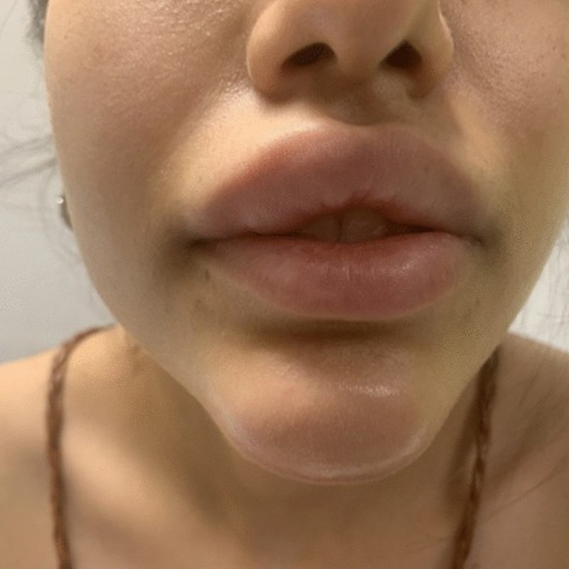
The core clinical patterns are recurrent edema, erythema, tenderness, induration, visible or palpable lumps, episodic inflammation and occasionally abscesses or ulceration. A clinician should document whether lesions are bilateral or asymmetric, whether there is one focal collection or multiple injection-site lesions, and whether the overlying skin is hot, discolored, intact or draining. Associated dental, sinus, skin or systemic infection, recent procedures, fever and prior filler history all affect the differential. [S1, S2]
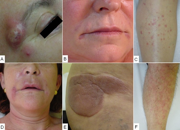
The multipanel case illustration in the source (Figure 1, originally around page 7) shows the diversity of appearances rather than a series of typical HA outcomes. Several panels involve older permanent or mixed materials, and another shows a distant skin inflammatory pattern. These photographs must never be promoted as before-and-after success images or as documentation of cases personally managed by the editorial physician. They are best interpreted with material identity and clinical history in mind. [S1]
Inflammatory versus noninflammatory lumps
A soft, painless, stable prominence near a previous injection site can represent gel accumulation or displacement rather than active immune inflammation. In contrast, increasing warmth, pain, redness and edema suggests an inflammatory lesion and raises the possibility of infection. A firm nodule may also be fibrosis or true granuloma. Ultrasound may help identify the material and morphology; aspiration or biopsy may be required in selected uncertain or persistent cases. The visible or palpable surface alone often does not establish the pathology. [S2, S5]
Why the lips and under-eye region deserve caution
Both sites contain mobile or delicate tissues where small deposits can become conspicuous. The lips are subject to repeated muscular motion, and the lower eyelids can make even small variations in water balance noticeable. Swelling may disproportionately influence appearance in these areas; this should not be mistaken for evidence that the underlying complication is always more dangerous. The appropriate response depends on vascular anatomy, original material, symptoms and imaging rather than appearance alone. [S1, S5]
7. A timeline of the cellular response: from healing to persistent inflammation
The source Figure 2 compares a stylized normal host response to a pathological, prolonged inflammatory reaction. In a typical resolving response, early innate-cell activity decreases as macrophages, fibroblasts and collagen remodeling progress. In a persistent reaction, inflammatory and fibrotic activity remains elevated for longer. This is a conceptual diagram, not a patient-specific prediction of what will happen at exactly one, four or eight weeks. The original figure is valuable because it frames late-onset granuloma as a host–material process evolving over time. [S1]
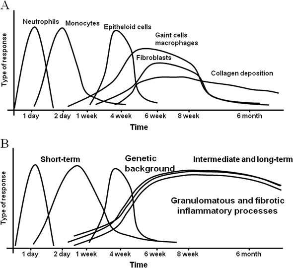
Under ordinary circumstances, immune surveillance and degradation/remodeling lead to tissue stabilization. Under some circumstances, inflammation continues or reactivates in the presence of retained material. Tissue persistence matters, which is why the old notion that a complication is impossible once the immediate healing period has passed is incorrect. However, long-lived product does not invariably lead to disease and ‘foreign body response’ is not automatically synonymous with ‘dangerous allergy.’ [S1, S3]
8. Pathogenesis: macrophages, granulomas, adjuvant hypotheses and biofilm debate
Granulomas are organized inflammatory structures often dominated by macrophages and multinucleated giant cells, with varying lymphocytes, fibroblasts and collagen. They represent the body’s attempt to contain material or an ongoing stimulus that it cannot readily clear. Fillers with differing chemistry and morphology may generate differing tissue patterns, but presentation is affected by host biology, microbiological factors, dose, location and time. The original review’s detailed immunopathology remains a useful background framework even though some causal ideas require modern qualification. [S1]
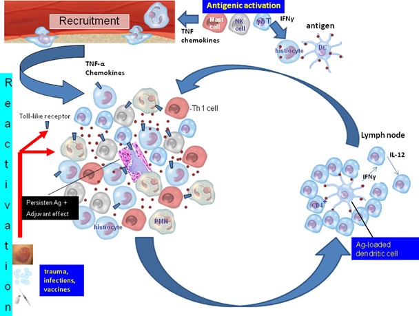
Are biofilms responsible? Microbial contamination and biofilm-like persistence have been proposed as contributors to delayed nodules. The literature is mixed: organisms can cause clear clinical infection, but in some unexplained inflammatory lesions routine cultures are negative and the role of a protected biofilm is difficult to demonstrate. The 2023 international consensus emphasizes the possible combination of filler, pathogens and an inflammatory host response; the 2024 CARE consensus explicitly considers filler physicochemistry, infection and immune imbalance as interacting hypotheses. Neither framework should be used to diagnose every late nodule as infection without evaluation. [S1, S2, S6]
What about systemic immune reactions? The old review includes case reports associating certain fillers and other biomaterials with sarcoid-like disorders, vasculitis, panniculitis and broader autoimmune phenomena. Case reports can identify a signal and justify further study; they do not independently establish a common causal risk. The proposed ASIA (autoimmune/inflammatory syndrome induced by adjuvants) construct is debated and should never be used to label a patient with autoimmune disease solely because they have filler or fatigue. Serious systemic symptoms warrant standard medical assessment and, when indicated, rheumatology, dermatology or other specialist input. [S1, S2]
9. What can biopsy and histopathology tell us?
When a lesion is persistent, atypical, progressive, recurrent or not explained by clinical examination and ultrasound, tissue pathology may help separate foreign-body granuloma from infection, sarcoid-like inflammation, neoplasm and other skin disease. Histology often includes multinucleated giant cells, epithelioid histiocytes, lymphocytes, fibrosis and product remnants. Pathologists may use material morphology, polarized light and special stains to suggest specific implant types, but products can overlap in appearance. A biopsy does not replace matching the pathology to the product history and clinical findings. [S1, S5]
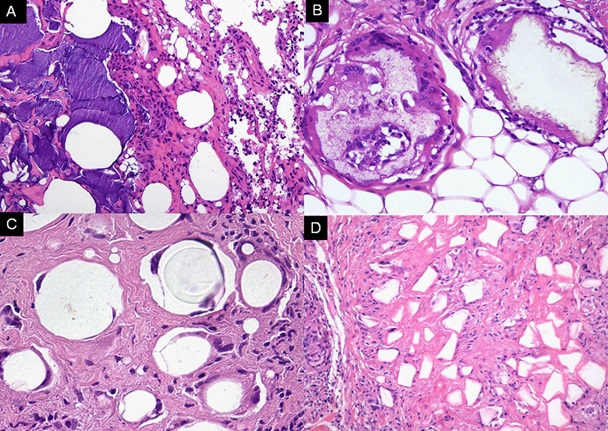
The 2013 source explains, for example, the characteristic empty-looking vacuoles around some nonresorbable oil-like materials and the recognizable rounded or polygonal structures in certain polymer implants. In HA-related lesions, glycosaminoglycan-rich material can appear basophilic and may be highlighted with Alcian blue staining. These patterns can suggest a class of product, but mixed prior injections, degradation and concurrent infection can complicate interpretation. A definite granulomatous diagnosis is histologic; many clinically described ‘granulomas’ in marketing discussions are simply unspecified nodules. [S1]
10. Diagnosis: a stepwise, individualized assessment
The first question is whether an emergency is present: skin ischemia, rapidly progressive infection, anaphylaxis, ocular symptoms, severe pain, fever or systemic instability cannot be managed as a routine cosmetic follow-up. For a stable patient, the clinician should construct a timeline: product name, batch or records if available, date, treatment sites, previous fillers and complications, onset, flare pattern, dental or other procedures, concurrent illness, medications and immune history. Important details also include prior attempts at dissolving or treating the lesion. [S2, S5]
| Finding | Possible explanation | Clinical next step |
|---|---|---|
| Soft stable painless lump | Filler deposit, migration, fibrosis | Records and examination; consider ultrasound |
| Tender red/hot nodule | Infection or sterile inflammation | Assess severity; consider ultrasound/sampling |
| Fluctuation / drainage / fever | Abscess or spreading infection | Prompt clinical review, cultures where feasible |
| Persistent atypical mass | Granulomatous or other pathology | Selected biopsy and specialist review |
| Pain, blanching, mottling, vision changes | Possible vascular emergency | Immediate emergency evaluation |
Examination should record symmetry, color, heat, tenderness, fluctuation, drainage, firmness, regional lymph nodes and signs in untreated areas. Standardized photography at rest and during expression can help document change. A skin abscess is not diagnosed by photographs alone; a stable palpable deposit is not an indication for indiscriminate antibiotics. Labs, targeted cultures or specialist assessment should follow clinical suspicion rather than a one-size-fits-all ‘filler blood panel’. [S2, S5]
High-frequency ultrasound, often with color Doppler, is increasingly valuable for confirming whether retained filler is present and identifying collections, migration, nodules, inflammatory thickening or altered local vascular findings. Sonography can distinguish some material signatures, although it is operator dependent and cannot provide a complete histologic diagnosis. Abscess suspicion may lead to image-guided sampling and microbiological testing. Biopsy is reserved for selected diagnostic uncertainty or persistent atypical disease. [S5, S7]
Interpreting ultrasound patterns carefully
HA deposits often appear relatively anechoic or hypoechoic; CaHA, PLLA, PMMA and silicone have other characteristic ultrasound patterns and artifacts that depend on formulation, timing and technique. An abscess may contain fluid with debris, while a granulomatous reaction can present as hypoechoic tissue surrounding filler deposits. Edema and cellulitis may cause diffuse tissue thickening. These features improve the differential diagnosis; they should not be treated as perfect sensitivity or specificity estimates in every setting. [S5]
The 2025 multinational ultrasound series analyzed 240 granulomatous-reaction cases involving multiple filler types. Because the sample was specifically selected from patients with granulomatous findings, the proportion of HA in that series is not the risk that an HA injection will cause a granuloma. This distinction is especially important for blogs discussing complication pictures: a clinic-based collection of complicated cases does not establish population incidence. [S7]
11. Clinical management principles: treat the diagnosis, not the label
| Working category | Evidence-informed approach | Caution |
|---|---|---|
| Noninflammatory HA accumulation | Assessment; selected hyaluronidase or observation | Confirm likely material; avoid automatic treatment |
| Inflammation without clear infection | Individual evaluation; observation or anti-inflammatory options | Do not suppress an unrecognized infection |
| Possible abscess/infection | Medical assessment, drainage/culture as indicated, antimicrobials | Hyaluronidase is not first-line infection therapy |
| Persistent granuloma/unknown material | Imaging, pathology, multidisciplinary care | Avoid uncritical repetition of old drug protocols |
A management plan must begin with a working diagnosis and a risk assessment. If a lesion is noninflammatory and represents misplaced or persistent HA gel, carefully targeted hyaluronidase may be an option under an appropriately trained clinician. If the problem is infection, drainage, microbiological assessment and antimicrobial management may be appropriate; the 2024 CARE board does not recommend hyaluronidase as the initial approach for active infection. Inflammatory lesions without evidence of infection may follow an individualized strategy that includes observation and, in selected circumstances, anti-inflammatory treatment. No reader should self-administer prescription medications based on this overview. [S2]
Inflammatory nodules require more nuanced decisions than simply ‘inject steroid’. Before suppressing inflammation, an infectious process needs to be considered, especially when there is warmth, progression, pus or systemic illness. Different algorithms in the literature place antibiotics, corticosteroids, hyaluronidase or observation at different points depending on the presumed etiology. These differences partly reflect the limited quality of comparative trials. The age of a protocol, the material injected and the local standard of care are essential context. [S1, S2, S6]
Hyaluronidase: useful primarily when an HA component is present and treatment of the implant itself is clinically justified. It does not dissolve CaHA, PLLA, PMMA or silicone. It can itself cause hypersensitivity, and dosing, timing, ultrasound targeting and use in potentially infected tissues require professional assessment. Emergency protocols for suspected HA-related vascular occlusion are separate from routine late-nodule care and must not be conflated. [S2, S5]
Antibiotics: appropriate for suspected or confirmed infection, guided by the clinical setting and cultures where relevant. Their use in purely inflammatory nodules without proven infection remains an area of differing expert practice; automatic prolonged or multidrug antibiotics can expose patients to side effects and resistance. An abscess may require drainage rather than oral medicine alone. [S1, S2]
Corticosteroids and immunomodulators: historically described for selected noninfectious inflammatory or granulomatous reactions, but they can worsen untreated infection, induce tissue atrophy and carry systemic risks. Intralesional agents, systemic steroids, methotrexate, calcineurin inhibitors, hydroxychloroquine, anti-TNF treatment and other therapies appear in old case literature; their inclusion in the source is not an endorsement of routine cosmetic-practice prescribing or a patient treatment recipe. Complex, persistent or systemic cases warrant appropriate specialists and consideration of the changing evidence base. [S1, S2]
Surgery or excision: sometimes considered for persistent residual material or refractory lesions, especially selected permanent implants. Operating in an actively inflamed region risks scarring, tissue injury and recurrence; a staged plan from a suitably qualified surgical and medical team is preferable to hasty removal. Most stable inflammatory problems require careful characterization before invasive intervention. [S1]
12. Red flags that should never be managed as an ordinary filler reaction
Call for emergency medical assessment when there is sudden severe or increasing pain, pallor, mottled or dusky skin, cold tissue, ulceration, new visual changes, facial weakness or signs of systemic compromise after a procedure. Sudden breathing difficulty, tongue/throat swelling, widespread hives with faintness or hypotension may indicate severe allergic reaction. Rapidly spreading redness, fever, confusion or a severely ill appearance raises concern for serious infection. These are warning patterns rather than a complete diagnostic checklist. [S5]
Do not delay urgent assessment while trying massage, applying skin products or waiting for routine clinic review. A clinician managing aesthetic injections should have documented escalation and emergency plans, familiarity with vascular and anaphylaxis protocols, and access to appropriate referral pathways. The present blog does not replace emergency training or a local institutional protocol. [S5]
13. Do vaccines, infections and dental procedures trigger filler swelling?
Some post-filler inflammatory episodes have been reported after viral or bacterial illness, vaccinations or dental procedures. The FDA recognizes that delayed swelling or redness near filler sites may follow such events. This is an association, not a blanket reason to avoid all vaccinations or medically necessary dental care, and not proof that every episode was caused by immune activation. Risk must be discussed individually. [S2, S5]
A 2024 expert group includes current health status and recent dental interventions among factors to consider when planning elective filler treatment; the group recommends postponing elective injections during active infections and maintaining careful aseptic technique. Blanket intervals for every vaccine, dental appointment or filler product cannot be derived from the older 2013 source. Patients should disclose any recent event and clinicians should avoid attributing symptoms to a single trigger without ruling out infection or other causes. [S2]
14. Autoimmune disease and filler suitability: avoid oversimplification
A history of autoimmune or inflammatory disease is part of informed risk assessment, not an automatic proof that an aesthetic procedure will provoke a flare. Disease activity, medication, immune suppression, prior reactions and the proposed filler type may affect decision-making. The 2013 review raises important rare systemic case descriptions but also acknowledges that epidemiologic evidence did not consistently establish a causal relationship in some contexts. Claims that ‘fillers definitely cause lupus’ or, conversely, ‘fillers cannot cause immune complications’ both overstate the evidence. [S1, S2]
For a patient with an established rheumatologic illness, active disease, immunosuppressive therapy or previously severe filler reaction, the treating aesthetic clinician should consider communication with the relevant specialist and individualized benefit-risk review. Alternative or deferral may be appropriate. Consent should distinguish reported associations, plausible mechanisms and demonstrated causal risk. [S1, S2]
15. Preventing avoidable complications before, during and after treatment
Prevention begins with accurate medical and product history, choice of licensed medical setting, verified and traceable material, anatomical knowledge, conservative planning and realistic goals. The patient should disclose previous products including those injected abroad, even if the exact brand is unknown. The physician should explain risks and alternatives, note site-specific indications, check active skin and dental infection, and record consent and product details. An older filler of unknown type should prompt caution before layering additional material into the same area. [S2, S5]
During treatment, meticulous asepsis, an appropriate sterile technique and correct anatomical placement reduce avoidable procedural and infectious risk. No injection approach—needle, cannula, superficial placement or a named signature technique—eliminates all vascular or delayed-inflammatory risks. After treatment, practical written instructions should explain expected recovery, when to contact the clinic, emergency symptoms and the importance of reassessment if swelling becomes recurrent or begins months later. [S2, S5]
Longitudinal documentation is a clinical asset. Preserving the product brand, lot, concentration, anatomical map, treatment date, relevant medication history and any past complication can make later diagnosis substantially more efficient. Good follow-up is especially important because a clinician who sees the patient years later may otherwise be unable to determine whether a lump is residual HA, a biostimulator, a permanent implant, fibrosis or another process. [S1, S5]
16. What patients in Abu Dhabi should ask at consultation
A practical consultation should establish the intended aesthetic outcome, the exact material and its reversibility, known and uncertain risks, which anatomical sites and planes are contemplated, the clinician’s emergency and follow-up arrangements, and the expected normal recovery timeline. Patients may also ask how the clinic handles problems arising long after injection or when the original product was used elsewhere. This is more helpful than judging safety by the brand name, price, number of syringes or an unqualified ‘zero risk’ promise. [S2, S5]
Readers can consult the related educational content at https://drerfanrahmani.com/services/dermal-filler-overview/ and product-class background at https://drerfanrahmani.com/services/juvederm-fillers/ or https://drerfanrahmani.com/services/calcium-filler-radiesse/. These are related pages for contextual reading, not endorsements that any brand is suitable for every patient. For an individual assessment, use the clinic contact page at https://drerfanrahmani.com/contact/. This article does not make claims about the availability of ultrasound, particular treatment equipment or specialist emergency services at any specific clinic without verification.
17. How to evaluate clinical evidence without being misled by dramatic cases
A vivid photograph illustrates what a reaction can look like, but says little about how often it occurs. A specialist center may publish patients with severe granulomas because those patients were referred there, while thousands of uneventful procedures are not represented. An expert consensus can help clinicians reason about uncommon problems but it is not a randomized trial proving that its algorithm is superior. A pooled rate may offer useful context but can hide variation in definitions, follow-up length and surveillance quality. [S1, S2, S3]
The historical source includes many reports of uncommon systemic disease; its chief value is mapping potential patterns and mechanisms rather than furnishing incidence estimates for today’s HA formulations. The September 2026 meta-analysis offers a newer quantitative perspective, yet incidence varies with study design and product technology, and it should not be applied mechanically to an individual consultation. The 2025–2026 ultrasound literature can improve diagnostic confidence, but it does not replace clinical assessment or pathology when uncertainty remains. [S1, S3, S5]
A practical evidence hierarchy for this subject
When available, well-conducted prospective comparative studies with meaningful follow-up generally provide stronger causal information than uncontrolled case series. Systematic reviews can synthesize those studies but inherit their limitations. Case reports are indispensable for detecting rare and unexpected events but cannot establish frequency. Consensus statements reflect expert interpretation and can differ where trials are scarce. Product labeling and current local professional standards matter directly for practice. [S2, S3, S5]
18. Conclusions: safety requires long-term thinking
The central message of the 2013 review remains valuable: implanted or injected biomaterials may produce both early and delayed adverse reactions, including immune-related inflammation, and complicated cases deserve thoughtful investigation. The modern update is equally important: available quantitative evidence suggests delayed inflammatory reactions to HA fillers are uncommon, but their timing is variable, their appearances overlap with infection and simple filler deposits, and no universal therapeutic recipe is supported for every presentation. [S1, S2, S3]
For patients, the most useful rule is to report unexpected, progressive or recurring swelling—even if the injection happened months ago—and seek immediate help for emergency symptoms. For clinicians, successful management depends on precise product history, differentiation between infection and sterile inflammation, ultrasound and sampling where indicated, individualized treatment, patient counseling and a clear referral pathway. In aesthetic medicine, a natural result and a responsible long-term safety plan are equally important. [S2, S5]
Frequently Asked Questions: inflammatory reactions to dermal fillers
Patient answers based on the evidence reviewed above.
1. Can hyaluronic acid filler cause inflammation months later?
Yes. Delayed reactions may begin weeks, months or occasionally years after HA filler placement. They can involve new swelling, redness, induration or tender nodules around a treated site. The key clinical task is to separate a sterile immune response from infection, residual product, migration or another disease rather than treating all delayed reactions as the same condition. A review of injection records and in-person assessment is recommended. [S2, S3]
2. Is a lump after filler always a granuloma?
No. A ‘lump’ is a symptom, whereas a granuloma is a specific organized tissue response, typically established histologically. Lumps can also represent product accumulation, superficial injection, fibrosis, infection, cysts or migration. Whether the lump is painful, inflamed, changing, or associated with other symptoms helps guide imaging and investigation. Avoid assuming that appearance alone proves granulomatous inflammation. [S1, S5]
3. How common are delayed inflammatory reactions to HA filler?
A September 2026 meta-analysis pooled an estimated 0.0005 events per patient-year across included studies, approximately one event per 2,000 patient-years of observation. This does not mean that every clinic, product or patient has the same risk; definitions and follow-up vary. It is also a rate per follow-up time, not an unconditional lifetime probability. [S3]
4. Can a reaction appear several years after treatment?
Yes, although it is unusual. The 2026 review’s case descriptions ranged from about two weeks to twelve years after injection, and the historical literature documents late reactions to a variety of filler materials. An old treatment history therefore remains relevant when investigating a new nodule, but timing alone never proves that filler caused the finding. [S1, S3]
5. Is filler swelling after a cold or flu an allergy?
Not necessarily. Intercurrent infection can coincide with or trigger swelling at an earlier filler site, but such episodes can be inflammatory without representing classical immediate allergy. The FDA recognizes swelling after some infections and viral illnesses. Assessment is needed if swelling persists, recurs, becomes painful or involves systemic symptoms, and infection must remain in the differential. [S2, S5]
6. Can dental treatment trigger a delayed filler reaction?
Temporal associations have been reported, and the 2024 consensus considers recent dental intervention among potential risk-related circumstances. However, the evidence does not justify claiming that every dental procedure causes filler inflammation. Patients should tell the dentist and aesthetic physician about relevant treatment histories and avoid elective aesthetic injections during active infection. Necessary dental care should not be delayed without medical advice. [S2]
7. Should I avoid vaccination if I have facial filler?
A universal ban is not supported by the evidence reviewed here. Transient inflammatory swelling near old filler sites has been described after some vaccinations, and the FDA lists vaccination among possible triggers. The significance depends on individual history and medical circumstances. Vaccination decisions should be made according to relevant public-health and treating-physician guidance, not fear generated by rare case reports. [S5]
8. Does filler cause autoimmune disease?
A direct causal relationship with broad autoimmune diseases has not been established simply by reporting cases after cosmetic injections. The 2013 review discusses rare systemic inflammatory presentations and proposed immune mechanisms, while noting that some epidemiologic data failed to confirm suggested associations. A patient with concerning systemic symptoms needs an ordinary medical evaluation, not an automatic diagnosis based on the presence of filler. [S1]
9. What is ASIA syndrome in filler discussions?
ASIA is a proposed autoimmune/inflammatory syndrome induced by adjuvants. It appears in historical biomaterial literature as a way of organizing certain symptoms and hypothesized immune effects, but its application to individual aesthetic patients is contested. It should not be diagnosed from filler history, fatigue or generalized symptoms alone; independent clinical evidence and appropriate specialist evaluation are essential. [S1]
10. Can filler become infected a long time after injection?
Late infection or inflammatory episodes suspected to involve microorganisms have been described, but the bacterial contribution is sometimes difficult to demonstrate. A new painful hot lesion, progressive erythema, draining material or systemic symptoms warrants prompt evaluation. Where appropriate, a clinician may arrange ultrasound, aspiration and cultures. Negative routine cultures do not automatically prove an immune-only mechanism. [S1, S2]
11. What is a filler biofilm?
A biofilm is a community of microorganisms protected within an extracellular matrix. Biofilms are established in some implanted medical devices; their exact role in all late facial filler nodules remains under investigation. Some expert models incorporate microbial persistence, while other lesions appear driven by sterile inflammation or product factors. ‘Biofilm’ is not a diagnosis that can be assigned to every red bump. [S1, S6]
12. What is the difference between swelling and infection?
Simple swelling can be soft, symmetric, minimally tender and progressively improving. Infection is more concerning when there is increasing heat, pain, focal tenderness, spreading redness, fluctuance, discharge or fever. The distinction is not absolute; imaging and clinical assessment may be necessary. A patient with worsening rather than settling symptoms should contact a qualified service, especially after an earlier period of improvement. [S2, S5]
13. Can a painless nodule be left alone?
Sometimes, particularly when it is stable, not inflamed and represents a harmless residual deposit. Nevertheless, new or persistent lumps deserve at least an assessment when the material is unknown, the lesion changes or the patient wants correction. Observation is a management decision following evaluation, not proof that every painless lesion is harmless. [S2]
14. Can hyaluronidase dissolve every kind of filler?
No. Hyaluronidase acts on hyaluronic acid; it is not a universal reversal enzyme for CaHA, PLLA, PMMA, silicone or many historic implant materials. The clinician should establish that HA is present and assess the risks and goal before using it. Non-HA materials may require entirely different approaches. [S1, S2]
15. Should hyaluronidase always be injected into an inflammatory nodule?
No. If infection is suspected, the 2024 CARE consensus advises against using hyaluronidase as an initial infection treatment because the infection needs its own evaluation and management. In a noninfectious HA deposit it can be useful; in a sterile inflammatory lesion different expert pathways may favor observation or anti-inflammatory treatment. The plan must be individualized. [S2]
16. Can hyaluronidase itself cause a reaction?
Yes. Hypersensitivity to hyaluronidase has been reported, although serious reactions are uncommon. The clinician should take the relevant allergy history and be prepared to manage adverse events. A product being used to resolve another injectable treatment’s complication does not make the second procedure automatically risk-free. [S1]
17. Are steroid injections safe for any filler granuloma?
Not universally. Intralesional steroids have been reported for selected inflammatory or granulomatous conditions, but they can cause skin atrophy, pigment change and other problems, and untreated infection can worsen with immunosuppression. Accurate diagnosis, material type, location and specialist experience matter. Older dosing suggestions in the 2013 paper should not be copied as a patient protocol. [S1, S2]
18. Do all filler nodules require antibiotics?
No. Antibiotics may be appropriate for bacterial infection and some expert pathways have considered them in uncertain delayed inflammatory nodules. They should not be automatically used for every noninfectious deposit, where the benefit is unproven and adverse effects and antimicrobial resistance remain relevant. A fluctuating collection may require drainage and cultures. [S2]
19. Can high-frequency ultrasound identify old filler?
Often it can help identify the location, distribution and likely material class, as well as swelling, granulomatous changes, collections or migration. HA and silicone, for example, tend to have different sonographic signatures. Results depend on device settings, operator skill, implant age and tissue conditions. An ultrasound is an adjunct to, not a replacement for, history, physical examination and selected pathology. [S5, S7]
20. Will ultrasound always distinguish granuloma from abscess?
No. Some features are suggestive, such as fluid with debris in an abscess or hypoechoic tissue surrounding filler in a granulomatous reaction, but overlap exists. Clinical history, examination, Doppler findings, sampling, microbiology and sometimes biopsy help resolve uncertain cases. The sonographic image alone should not trigger a lengthy immunosuppressive or antibiotic course without a defensible diagnosis. [S5]
21. When is biopsy considered after filler?
Biopsy may be appropriate for an unexplained, persistent, atypical or recurrent mass, particularly when infection, foreign-body reaction and another dermatologic condition cannot be separated clinically. Tissue examination may identify macrophage and giant-cell reactions, fibrosis or material-specific clues. Decisions are individualized because facial biopsy can create scarring and may not be necessary for straightforward cases. [S1]
22. Why do clinicians request the brand and injection date?
Different filler families have different persistence, imaging patterns and reversal options. Exact brand, batch, volume, site and timing allow the clinician to distinguish a likely HA deposit from a product that does not respond to hyaluronidase. Missing records increase uncertainty; this is one reason to keep copies of treatment details even long after a successful aesthetic procedure. [S1, S5]
23. Is HA filler always safer than a permanent filler?
HA is widely used and has the practical advantage of biodegradability and potential enzymatic reduction, while some permanent materials have historically caused very difficult long-term reactions. However, no injected material is completely without risk, and safety depends on anatomy, product labeling, patient factors and technique. ‘Reversible’ should never be interpreted as ‘guaranteed safe’. [S1, S5]
24. Can Radiesse cause a granuloma?
Foreign-body inflammatory reactions and nodules have been reported after calcium hydroxylapatite products. Risk and management vary by area, technique and clinical setting. CaHA is not HA, so standard hyaluronidase does not dissolve it. Product selection and appropriate clinical indication are especially important in mobile or delicate anatomical sites. [S1]