
Meta Cell Technology in Abu Dhabi: complete guide
What Are Autologous PRP-Derived Exosomes ?
Exosomes are nanoscale extracellular vesicles that help cells communicate. The study describes them as approximately 30–150 nm structures surrounded by a lipid bilayer and carrying proteins, nucleic acids and other biologically active molecules. They are produced by many cell types and can be found in body fluids including blood, saliva, urine, cerebrospinal fluid and milk.
In regenerative medicine, exosomes are of interest because they can transport signaling molecules between cells. The paper specifically focuses on exosomes associated with platelet-rich plasma rather than externally manufactured or donor-derived exosome products.
Platelets contain and release growth factors, cytokines, chemokines and extracellular vesicles. The source notes that platelet-derived exosomes can also carry microRNAs, which are small non-coding RNAs involved in regulating gene expression. The broader literature cited in the paper links platelet-derived extracellular vesicles to processes such as angiogenesis, inflammation, wound healing, apoptosis and cell growth.
Why PRP Is Relevant to Regenerative Facial Treatment
The article places PRP within personalized medicine because it is autologous: the biological material originates from the patient’s own blood. The authors describe PRP as accessible and capable of releasing endogenous growth factors that resemble the body’s own repair signals.
The central concept of the study is not simply to inject standard PRP. Instead, the PRP is processed using a photothermal-biomodulation protocol intended to precondition platelets and promote the release of platelet-derived exosomes before facial administration.
What Is Photothermal Biomodulation in This Study ?
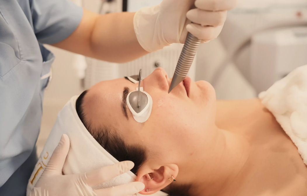
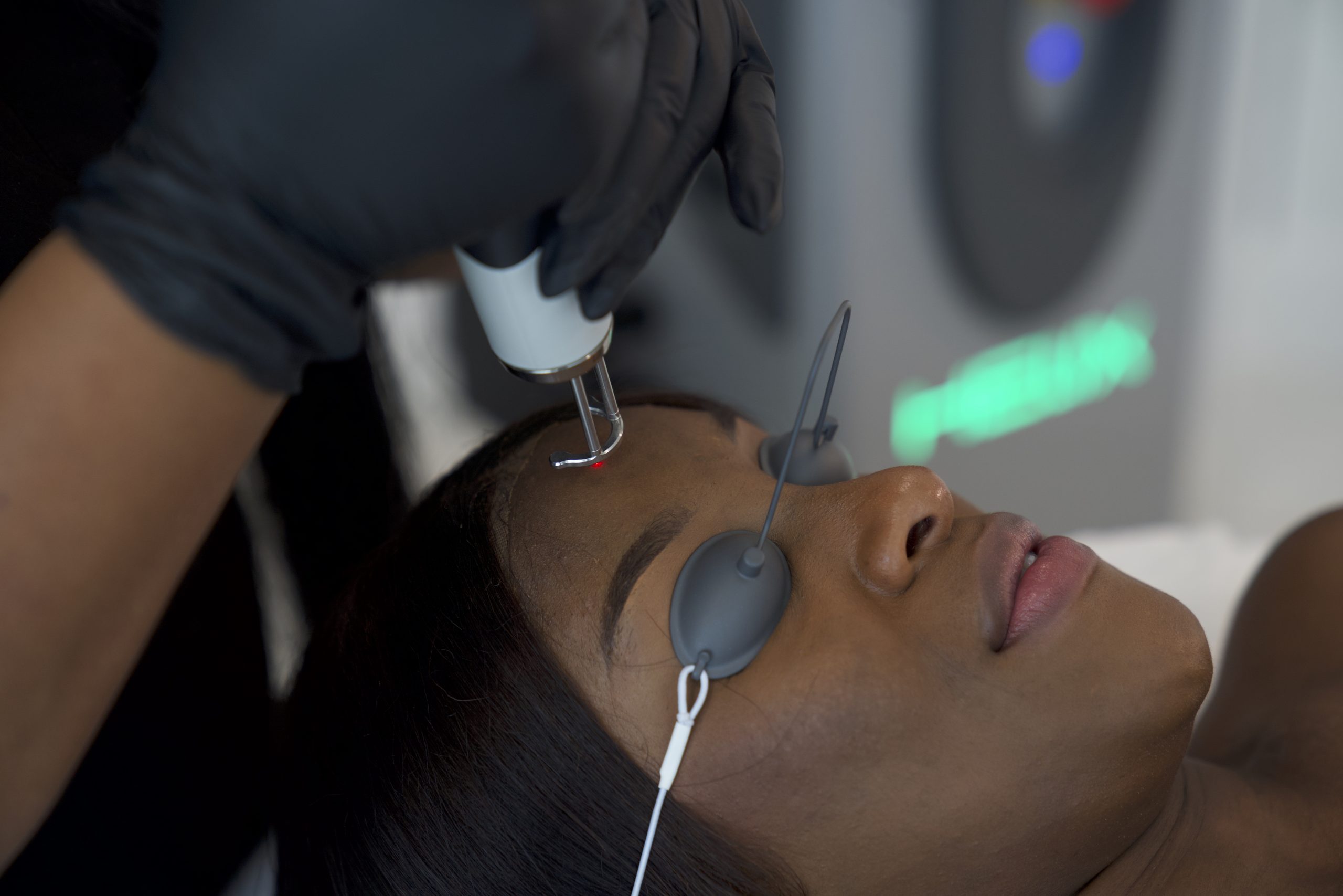
The Meta Cell Technology System is described as a device with predefined programs combining light energy, wavelength, temperature and exposure time. The aim is to precondition autologous cells or platelets in a reproducible way.
For this facial-rejuvenation pilot, the investigators used the system’s ‘Exosomes’ preset. The source reports 467 nm light at 2 J/cm² while maintaining the sample at 37°C for 10 minutes.
A Split-Face Randomized Design — With an Important Interpretation Caveat
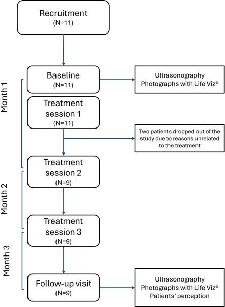
The study states that half of the face was randomly assigned to receive the autologous PRP-derived exosome treatment. Baseline ultrasound measurements were obtained on both sides of the face to confirm that the sides were comparable before treatment.
However, the numerical follow-up table presented in the paper focuses on the treated side, and the main published efficacy figures are changes from baseline on that treated side. For that reason, this article does not overstate the paper as definitive proof of superiority over the untreated side.
Who Was Excluded From the Study ?
| Exclusion criterion | Source description |
|---|---|
| Pregnancy or breastfeeding | Excluded |
| Malignancy | Excluded |
| Viral infections | Hepatitis B, hepatitis C, HIV or syphilis |
| Systemic autoimmune disease | Excluded |
| Hematologic disease | Excluded |
| Recent facial rejuvenation | Prior rejuvenation treatment within 6 months |
| Immunosuppressant therapy | Excluded |
| Anticoagulant therapy | Excluded |
These criteria define the study population; they do not automatically constitute a universal contraindication list for every future PRP or exosome-related protocol. Current device instructions, local regulation and patient-specific medical assessment remain necessary.
How the Product Was Prepared
For each session, the source reports collection of 10 mL of peripheral blood into citrated tubes. The sample was centrifuged at 1800 rpm for eight minutes and processed using a closed commercial PRP system.
The paper states that this process yielded approximately 4–5 mL of PRP. It then describes transferring 8–10 mL of PRP into the MCT Kit before applying the ‘Exosomes’ preset.
Internal Source InconsistencyThe methods text contains a volume inconsistency: it reports a PRP yield of approximately 4–5 mL and then states that 8–10 mL of PRP were transferred to the MCT Kit. The paper does not reconcile this discrepancy, so this guide does not silently correct it.
Injection Technique Used in the Pilot Study
The treated half-face was cleansed with chlorhexidine. A topical lidocaine-prilocaine anesthetic cream was applied 45 minutes before injection.
The source reports multipuncture mesotherapy using a 30-gauge needle at approximately 0.5 cm intervals, with the needle positioned at about a 30-degree inclination. Each puncture delivered 0.1 mL at an approximate depth of 2.0 mm.
Each treatment visit lasted approximately one hour per patient. These details belong to this specific research protocol and should not be interpreted as a universal recipe for another device, product preparation or patient.
Ultrasound Results : The Strongest Numerical Finding
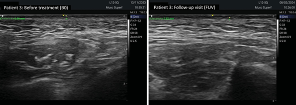
One month after the third session, mean SLEB thickness on the treated side decreased substantially. The cheekbone showed a mean reduction of 61.39%, while the cheek showed a mean reduction of 39.35%.
The source reports a cheekbone reduction range of approximately 51.0% to 71.8% and a cheek range of approximately 23.8% to 55.0% in the summary text. Individual values in Table 1 vary more widely, particularly in the cheek.
| Patient | Age | Cheekbone baseline (mm) | Cheekbone follow-up (mm) | Change | Cheek baseline (mm) | Cheek follow-up (mm) | Change |
|---|---|---|---|---|---|---|---|
| 1 | 55 | 1.10 | 0.61 | -44.55% | 0.90 | 0.54 | -40.00% |
| 2 | 51 | 1.70 | 0.67 | -60.59% | 1.60 | 0.59 | -63.13% |
| 3 | 57 | 1.30 | 0.40 | -69.23% | 0.90 | 0.52 | -42.22% |
| 4 | 65 | 1.20 | 0.36 | -70.00% | 1.30 | 0.59 | -54.62% |
| 6 | 56 | 1.20 | 0.64 | -46.67% | 1.30 | 0.72 | -44.62% |
| 7 | 56 | 1.30 | 0.59 | -54.62% | 1.10 | 0.63 | -42.73% |
| 9 | 55 | 1.20 | 0.37 | -69.17% | 1.00 | 0.66 | -34.00% |
| 10 | 62 | 1.00 | 0.29 | -71.00% | 0.80 | 0.64 | -20.00% |
| 11 | 56 | 0.90 | 0.30 | -66.67% | 0.70 | 0.61 | -12.86% |
| Average | 57.0 | 1.21 | 0.47 | -61.39% | 1.07 | 0.61 | -39.35% |
How Should SLEB Reduction Be Interpreted ?
The study interprets the reduction in SLEB thickness as evidence of improvement in the ageing-related subepidermal band. The authors connect the finding to proposed exosome effects on fibroblasts, collagen fiber density and dermal regeneration.
The study did not perform skin biopsy or direct histologic collagen quantification. Therefore, the safest wording is that ultrasound changes were consistent with dermal improvement rather than proving a specific amount of new collagen formation.
LifeViz 3D Skin Analysis
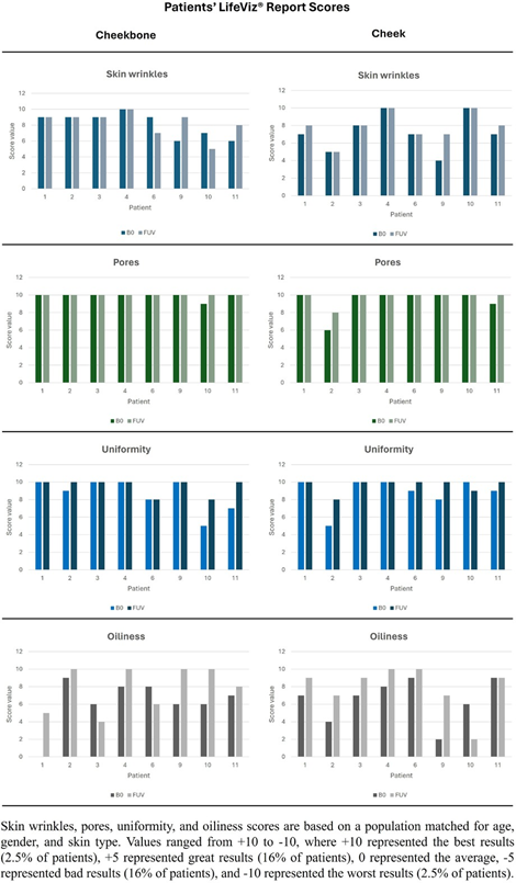
The investigators also used the LifeViz Mini stereophotogrammetric 3D imaging system. It generated scores for wrinkles, pores, skin uniformity and oiliness relative to a population matched for age, gender and skin type.
Photographic imaging data were available for eight patients rather than all nine completers. This matters when interpreting percentages because the denominator for LifeViz outcomes was often eight.
Wrinkles, Pores, Uniformity and Oiliness — What Changed ?
The digital skin results were heterogeneous rather than uniformly positive. Oiliness was the parameter with the greatest number of patients showing improvement, while pores most often remained unchanged.
The paper reports that 50% of photographed patients showed improvement in oiliness in both assessed regions. For pores, 87.5% showed no change at the cheekbone and 75% showed no change at the cheek.
The discussion summarizes LifeViz improvement rates across measured features as ranging from 25% to 75% of patients, with the most pronounced effects in oiliness, followed by uniformity, pores and wrinkles.
| LifeViz parameter | Cheekbone average change | Cheek average change | Key interpretation from source |
|---|---|---|---|
| Wrinkles | +4.1% | +12.9% | Variable; some patients improved, others did not |
| Pores | +1.4% | +5.6% | Most patients showed no change |
| Uniformity | +14.2% | +12.2% | Improvement in a subset of patients |
| Oiliness | +17.9% | +43.9% | Most frequently improved parameter |
Patient-Reported Outcomes
Seven of nine completers (77.8%) said their skin had improved one month after the final session. Four patients (44%) reported brighter skin.
Other reported changes included smoother skin, improved texture, fewer wrinkles, reduced sagging and less prominent eye wrinkles. Two patients (22.2%) said they did not perceive a difference.
The discussion section later describes 75% of patients as reporting improved skin quality. Because the explicit results section gives a numerator and denominator of 7/9, this guide uses 77.8% as the primary figure and notes the inconsistency rather than silently reconciling it.
| Patient-reported change | Study result |
|---|---|
| Any perceived skin improvement | 7/9 (77.8%) |
| Brighter skin | 4/9 (44%) |
| Smoother skin | 1/9 (11.1%) |
| Improved skin texture | 2/9 (22.2%) |
| Reduced wrinkles | 1/9 (11.1%) |
| Reduced facial sagging | 2/9 (22.2%) |
| Less prominent eye wrinkles | 1/9 (11.1%) |
| No perceived difference | 2/9 (22.2%) |
Representative Before-and-After Images
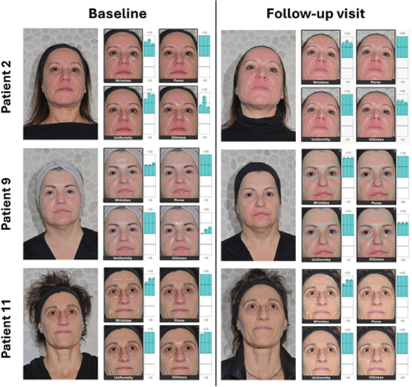
The source includes representative baseline and follow-up images for patients 2, 9 and 11. The follow-up images were obtained one month after the final treatment session.
The figure combines standard photographs with LifeViz analysis for wrinkles, pores, uniformity and oiliness. These examples help visualize the type of change seen in selected participants, but they should not be treated as a guarantee of the same result in every patient.
Safety and Adverse Events
The treatment was generally well tolerated in the study. One of nine completers (11.1%) reported swelling with bruising after the first session.
The same patient had less swelling and inflammation during the second and third sessions. The paper concludes that no significant adverse effects were associated with the treatment.
The small cohort means uncommon adverse effects could easily have been missed. ‘Well tolerated in this pilot study’ is therefore more defensible than ‘risk-free.’
What the Study Suggests About Skin Biology
The authors link the findings to the proposed cellular effects of platelet-derived exosomes. In the biological model described by the paper, exosomes may activate fibroblasts, influence collagen fiber density and support dermal regeneration.
The cited literature also associates platelet-derived extracellular vesicles with inflammation modulation, wound healing, angiogenesis and cell survival. These mechanisms provide biological plausibility for rejuvenation, but the clinical pilot itself did not isolate a single molecular mechanism responsible for the visible changes.
What Does ‘Autologous Exosomes’ Mean Here ?
The term ‘autologous’ means the biological material came from the same patient. This distinguishes the protocol from externally manufactured, donor-derived or cell-culture-derived exosome products.
In this study, the treatment material began as the patient’s own blood, was processed into PRP and then photothermally preconditioned with the MCT system. The source describes the final treatment as autologous PRP-derived exosomes.
This terminology is important for website accuracy. It is better to describe exactly what was studied than to use the broad label ‘exosome facial’ without identifying the biological source and preparation method.
What the Study Supports
- Three monthly sessions of the source-specific autologous PRP-derived exosome protocol were feasible in this small cohort.
- Nine patients completed the study and objective ultrasound follow-up.
- SLEB thickness decreased by an average 61.39% at the cheekbone and 39.35% at the cheek on the treated side.
- LifeViz analysis showed variable changes in wrinkles, pores, uniformity and oiliness.
- Oiliness was the parameter with the most frequent improvement in the photographed subgroup.
- Seven of nine participants reported overall skin improvement.
- One patient reported swelling and bruising after the first session.
- The treatment was described as well tolerated overall in the pilot study.
What the Study Does Not Prove
- That every patient will achieve a 61% or 39% SLEB reduction.
- That all wrinkle, pore, uniformity and oiliness scores improve in every patient.
- That PRP-derived exosomes are superior to standard PRP, skin boosters, laser, RF, microneedling or other rejuvenation treatments.
- That the visible changes are durable beyond the short follow-up period.
- That the same results apply to men or younger patients, because all completers in this study were women aged 51–65.
- That a precise therapeutic exosome dose was quantified in each patient.
- That absence of significant adverse effects in nine completers proves a zero-risk profile.
Important Limitations
The authors explicitly acknowledge several limitations: small sample size, follow-up limited to one month after the final session, and lack of directly comparable studies using this specific autologous PRP-derived exosome approach.
Although the paper describes a randomized controlled design, the main numerical follow-up data presented in the article focus on treated-side change from baseline. The article does not provide a large powered comparison capable of establishing superiority or long-term maintenance.
The cohort was also narrow: all participants were women and the mean age was 57 years. A larger and more diverse sample would be needed before generalizing these findings to all facial-rejuvenation patients.
Conflict-of-Interest and Evidence-Integrity Context
The disclosure section states that Laura Cordero, PhD, and Hernán Pinto, PhD, were employees of the scientific department of Meta Cell Technology. The remaining author declared no conflict of interest.
Employment by the device company does not invalidate the findings, but it is relevant context when interpreting a small device-specific pilot study. Independent replication would strengthen confidence in the results.
The reference list also contains at least one cited article that was later retracted. That reference is not central to the primary clinical outcome data in this pilot, but it is another reason to evaluate mechanistic background literature critically.
Who Might Be Worth Assessing for This Type of Treatment ?
The source studied adults with visible facial ageing signs rather than one narrowly defined wrinkle or area. The measured outcomes included skin structure, wrinkles, pores, uniformity, oiliness and subjective appearance.
A real-world consultation should first identify what the patient wants to improve. Fine lines, texture, pore appearance, skin quality, laxity and volume loss are different problems and may not respond equally to a regenerative injectable approach.
The study excluded pregnancy, breastfeeding, malignancy, several viral infections, systemic autoimmune or hematologic disease, recent rejuvenation treatment and use of immunosuppressants or anticoagulants. Medical eligibility should therefore be assessed individually.
How This Fits Into a Broader Facial-Rejuvenation Plan
This study does not compare autologous PRP-derived exosomes directly with botulinum toxin, dermal filler, biostimulators, energy-based devices, standard PRP or skin boosters.
That is clinically important because these treatment categories address different biological targets. A patient whose main concern is dynamic muscle activity, structural volume loss or severe laxity may need a different strategy than a patient seeking gradual improvement in skin quality.
For an evidence-based treatment plan, regenerative injectables should be considered within the context of diagnosis rather than selected solely because the term ‘exosome’ is popular.
Facial Rejuvenation with Autologous PRP-Derived Exosomes in Abu Dhabi
For patients searching for PRP-derived exosome facial rejuvenation in Abu Dhabi, the central question is whether a regenerative skin-quality approach matches the patient’s actual goals and medical profile.
Dr. Erfan Rahmani can assess skin quality, laxity, fine lines, prior procedures and the relative role of injectable versus device-based treatment before deciding whether a regenerative protocol is appropriate.